
Epidemiological data of pregnant women with fetuses or newborns diagnosed with amniotic band syndrome in Brazil
Authors:
Y. C. Da Silva Neri 1; E. Araujo Júnior 2,3; R. M. Moreira 1; L. M. Teixeira 3; L. M. de Ávila 1
Authors‘ workplace:
Department of Obstetrics and Gynecology, University of Vassouras, Vassouras – RJ, Brazil
1; Department of Obstetrics, Paulista School of Medicine – Federal University of São Paulo (EPM-UNIFESP), São Paulo – SP, Brazil
2; Discipline of Woman Health, Municipal University of São Caetano do Sul (USCS), São Caetano do Sul – SP, Brazil
3
Published in:
Ceska Gynekol 2026; 91(3): 193-197
Category:
Original Article
doi:
https://doi.org/10.48095/cccg2026193
Overview
Objective: This study aimed to analyze epidemiological aspects in Brazil related to women who had fetuses or newborns diagnosed with amniotic band syndrome (ABS), to assess perinatal outcomes of the newborns who received this diagnosis, and to examine the patients’ perception regarding the knowledge and support provided by healthcare professionals involved at the time of diagnosis. Methods: This was a quantitative, descriptive, analytical, retrospective, and cross-sectional study based on an online questionnaire administered at a single time point. The sample was randomly defined and included voluntary participation of female patients with a history of pregnancy affected by ABS. Results: Twenty-two questionnaires were analyzed. There was a predominance of cesarean deliveries and miscarriages among women with a history of ABS. ABS most frequently affected the upper limbs, particularly the hands and arms. A healthcare professional made the diagnosis during prenatal care via ultrasonography in 54.5% of the cases; however, only 40.9% of professionals were reported to be well-informed and to have provided full support. Fetal surgery was performed in only one case diagnosed during prenatal care. Cesarean delivery was predominant, as reported by 17 participants. Out of the 21 children who survived, 54.5% weighed > 2,500 g. Conclusion: ABS should receive greater emphasis during medical education and professional training updates to enable early diagnosis, intrauterine surgical treatment when indicated, and – most importantly – to ensure adequate guidance and support to the families.
Keywords:
amniotic band syndrome – amnion – intrauterine amputation – intrauterine constriction ring
Introduction
Amniotic Band, also known as constriction rings, refers to situations in which amniotic tissue – the inner lining of the amniotic sac – detaches and begins to float in the amniotic fluid surrounding the fetus [1]. These floating bands may lead to amniotic band syndrome (ABS), a set of congenital malformations caused by the entanglement of fetal body parts by constriction rings, disrupting normal development and potentially resulting in miscarriage [2]. Treatment can be conservative or surgical, depending on the type and severity of the anomaly [3].
ABS is a non-genetic condition that occurs sporadically in nature. Epidemiological data indicate an incidence ranging from 0.19 to 8.1 per 10,000 births, affecting male and female infants equally [4]. Major risk factors include high altitude (causing hypoxia), black ethnicity, primiparity, young maternal age, obesity, drug abuse during pregnancy (particularly cocaine), smoking, uterine malformations, acute febrile illness, 1st-trimester bleeding, collagen vascular disease, and previous uterine surgery. Diabetic vasculopathy may also increase the risk of amniotic rupture [5,6].
A wide spectrum of clinical deformities may occur in ABS cases, ranging from simple constrictions to severe craniofacial or visceral defects [7]. The syndrome is frequently associated with digital or limb amputations, syndactyly, acrosyndactyly, and malformations of the face, thorax, or abdomen. The extremities are most commonly affected, with distal regions of the upper limbs – especially the central fingers (II, III, and IV) – most frequently involved. In the lower limbs, the hallux and second toe are most often affected [8].
Early diagnosis of ABS can be achieved at the end of the 1st trimester through ultrasound examination, which allows visualization of limbs with restricted movement or altered blood flow. However, the mere ultrasonographic presence of amniotic bands does not qualify for an ABS diagnosis in the absence of concurrent fetal malformations [9]. Indications for intrauterine surgical intervention are based on the presence of neurological, vascular, or lymphatic compromise. When indicated, intrauterine surgery consists of releasing the constricting amniotic bands – often through Z-plasties – to restore skin and subcutaneous tissue length [10].
In this context, it’s relevant to emphasize the need for early detection of ABS, a condition that, although rare, can have significant consequences for fetal development and postnatal quality of life. It is also important to assess diagnostic factors, raise awareness about the condition, and encourage collaboration among healthcare professionals to ensure optimal care and family support. The present study aimed to analyze epidemiological data related to patients who had fetuses or newborns diagnosed with ABS in Brazil. As secondary objective, we proposed to analyze patients’ perceptions regarding the knowledge of health professionals who assisted them at the time of diagnosis.
Methods
This was a descriptive, analytical, retrospective, and cross-sectional study using a quantitative approach and random sample selection. The research was disseminated primarily through social media (Instagram, Meta Platforms Inc., Menlo Park, CA, USA), especially on the page of the “Associação Dar a Mão,” which has approximately 10,000 followers, many of whom are mothers of children affected by ABS. The questionnaire was made available online through a link, and any woman meeting the inclusion criteria could participate voluntarily after signing the informed consent form. Participants could withdraw at any time by not completing the form.
Inclusion criteria comprised of women aged 18 or older, with a history of pregnancy affected by ABS, and with internet access. The exclusion criteria encompassed women without a history of ABS or without internet access.
The form (https: //forms.gle/p2mDUKu6zmyciTom9) was designed to identify physiological characteristics associated with the occurrence of ABS and to assess the diagnostic process for these women. Completed forms were automatically sent to the researchers, who organized the data into Excel 2010 spreadsheets (Microsoft Corp., Redmond, WA, USA) for quantitative analysis through graphs and tables.
This study aims to contribute to the scientific community by expanding knowledge about early diagnosis, family counseling, and appropriate management of ABS. It also seeks to provide the general population with information about a condition that can be diagnosed during pregnancy and better managed through appropriate treatment. Risks include potential recall bias, data exposure, and data loss, which were minimized through secure data storage and confidentiality as stated in the informed consent form. All collected data were used exclusively for research purposes, ensuring participant confidentiality.
The project was approved by the Research Ethics Committee of Vassouras University under CAAE number 83965924.5.0000.5290. All participants signed the consent forms.
Results
The online questionnaire yielded 22 responses, with all participants agreeing to the informed consent and confirming a diagnosis of ABS during pregnancy or the postpartum period.
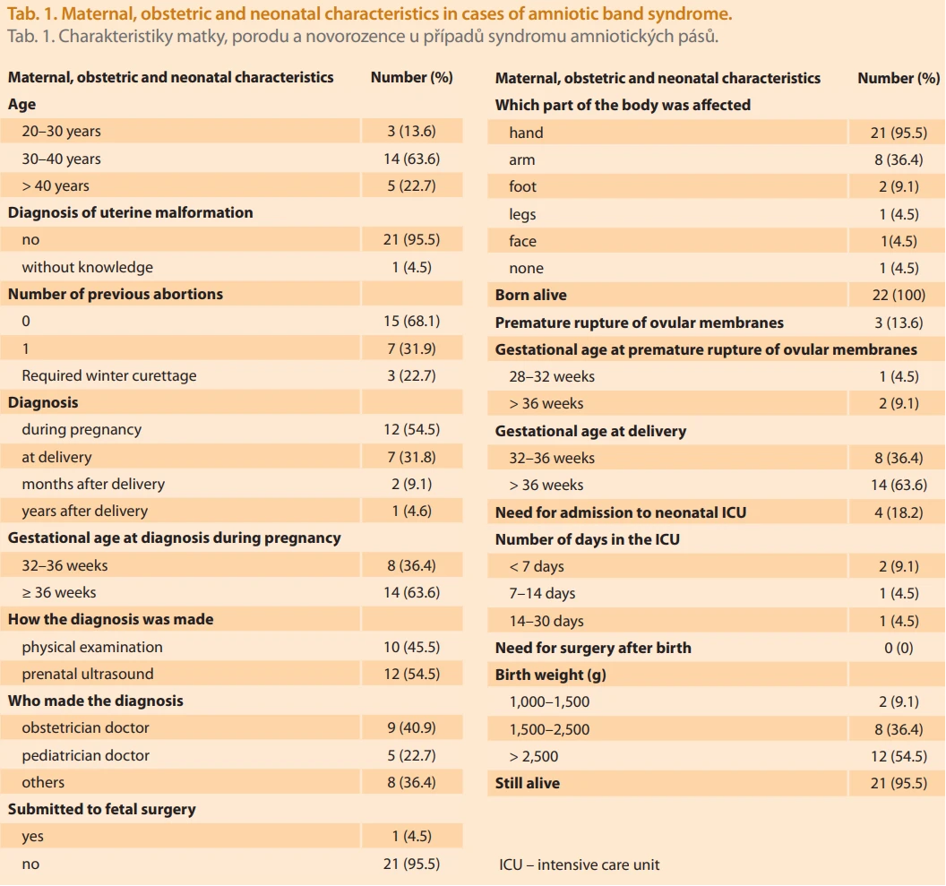
Regarding pregnancy history, 50% (11 women) had more than two pregnancies, 27% (6 women) had one pregnancy, 13.6% (3 women) had three, and 9.1% (2 women) had four; none had more than five. Cesarean delivery was predominant, reported by 17 participants (9 with one cesarean, 6 with two, and 2 with three). Vaginal delivery occurred in 9 women (5 with one vaginal delivery, 2 with two, and 2 with three).
Among the total number of women, eight participants (36.4%) reported miscarriages – seven of those before the pregnancy affected by ABS and one after the pregnancy affected by ABS. Out of the patients who suffered miscarriage, five required curettage procedures. Regarding premature rupture of ovular membranes, 86.4% did not experience it, while 13.6% reported fluid loss before delivery (9.1% after 36 weeks, 4.5% between 28 and 32 weeks).
Most participants were aged 30–40 years (14 women), followed by those over40 (5 women) and 20–30 years (3 women). None were under 20 years old. A total of 95.5% reported no uterine malformation diagnosis, and 4.5% were unsure. Deliveries of pregnancies affected by ABS occurred after 36 weeks in 14 cases and between 32 and 36 weeks in 8 cases. Only 54.5% (12 women) received the diagnosis prenatally through ultrasound performed by a healthcare professional; none underwent magnetic resonance imaging. The remaining 45.5% received the diagnosis postnatally, either visually at delivery or by a healthcare provider. Additionally, 9.1% were informed about the diagnosis by relatives or friends after birth. Diagnosis was communicated by an obstetrician in 40.9% of cases, by a pediatrician in 22.7%, and by another healthcare professional in 36.4%. Fetal surgery was performed in only one case diagnosed during prenatal care.
Regarding postnatal outcomes, 40.9% of mothers described their child’s health as good with no limitations, 45.5% as good with minor limitations, and 9.1% as good with significant limitations. The newborns were evenly distributed by sex (50% male, 50% female). Twenty-one survived; one died two hours after birth. Of the 21 children who survived, 54.5% weighed > 2,500 g, 36.4% weighed between 1,500 and 2,500 g, and 9.1% between 1,000 and 1,500 g. The most frequently affected organ was the hand (21 cases), followed by the arm (8 cases) and foot (2 cases). None underwent postnatal surgery.
After birth, 18.2% (4 infants) required intensive care unit admission – two for over one year, one for 14–30 days, and one for 7–14 days. Among those diagnosed by a healthcare professional, 40.9% reported that the professional was well-informed and provided full support, whereas 27.3% stated that not all questions were adequately addressed. Tab. 1 summarizes the main results of epidemiological data of ABS cases.
Discussion
Amniotic band syndrome is a rare condition associated with amniotic rupture, leading to the formation of fibrous strands during pregnancy. These may constrict fetal limbs, compromise the umbilical cord, cause malformations, and – in severe cases – pose a risk to fetal life [11]. Diagnosis is achieved by two - or three-dimensional ultrasound, magnetic resonance imaging, or postnatal clinical presentation [12].
The extremities are the most frequently affected regions, often showing constriction rings that can cause lymphedema, vascular and neural impairment, as well as aesthetic alterations. Cranial, facial, thoracic, and abdominal manifestations may also occur, though less commonly [13]. Prognosis depends on the range of manifestations, and follow-up is performed through obstetric ultrasonography and fetal echocardiography [12].
Because of the wide spectrum of clinical presentations, epidemiological data on ABS are difficult to specify [12]. No significant differences are observed between sexes. The incidence is markedly higher among stillbirths (110 per 1,000), possibly due to severe umbilical cord constriction [14].
There are no standardized guidelines for the management of ABS; clinical monitoring must be individualized, with appropriate counseling to pregnant women regarding detected and potential undetectable anomalies [12]. Surgical treatment, involving the release of amniotic bands, depends on the affected organ’s location and functionality.
Surgical procedures are more frequently indicated when vascular and/or neural compromise is present, allowing partial or total recovery of the affected area [15]. Intrauterine fetoscopic intervention, indicated in cases of high fetal mortality risk or severe impairment, may restore blood flow, delay constriction, and increase the chances of preserving limbs or preventing stillbirth, taking advantage of fetal tissue plasticity [10,12].
Conclusion
In summary, there was a predominance of cesarean deliveries and miscarriages among patients with a history of ABS. The syndrome most frequently involved the upper limbs, particularly the hands and arms. Only 54.5% received the diagnosis prenatally by ultrasound performed by a healthcare professional, and just 40.9% of these professionals were considered well-informed and supportive. Given its rarity and potential severity, ABS should be more thoroughly addressed in undergraduate medical education and continuing professional training, to enable early diagnosis and provide appropriate guidance and support to affected families.
Sources
1. Society for Maternal-Fetal Medicine; Gandhi M, Rac MW, McKinney J. Amniotic band sequence. Am J Obstet Gynecol 2019; 221 (6): B5–B6. doi: 10.1016/j.ajog.2019.09.020.
2. Sobana M, Halim D, Faried A et al. Amniotic band syndrome with CNS involvement: a pediatric neurosurgeon’s dilemma-a case series and literature review. Childs Nerv Syst 2023; 39 (8): 2003–2017. doi: 10.1007/s00381-023-06008-w.
3. Komarčević A, Pajić M, Djermanov M et al. Single stage surgical treatment of amniotic band syndrome – case report. Srp Arh Celok Lek 2016; 144 (9–10): 545–549.
4. López-Muñoz E, Becerra-Solano LE. An update on amniotic bands sequence. Arch Argent Pediatr 2018; 116 (3): e409–e420. doi: 10.5546/aap.2018.eng.e409.
5. Fatema N, Acharya Y, Yaqoubi H. Amniotic band ayndrome: a silent knife in-utero. Nepal Med Coll J 2019; 21 (2): 153–159. doi: 10.3126/nmcj.v21i2.25116.
6. Cignini P, Giorlandino C, Padula F et al. Epidemiology and risk factors of amniotic band syndrome, or ADAM sequence. J Prenat Med 2012; 6 (4): 59–63.
7. Walter JH Jr, Goss LR, Lazzara AT. Amniotic band syndrome. J Foot Ankle Surg 1998; 37 (4): 325–333. doi: 10.1016/s1067-2516 (98) 80070-7.
8. Moran SL, Jensen M, Bravo C. Amniotic band syndrome of the upper extremity: diagnosis and management. J Am Acad Orthop Surg 2007; 15 (7): 397–407. doi: 10.5435/00124635-200 707000-00005.
9. Hata T, Tanaka H, Noguchi J. 3D/4D sonographic evaluation of amniotic band syndrome in early pregnancy: a supplement to 2D ultrasound. J Obstet Gynaecol Res 2011; 37 (6): 656–660. doi: 10.1111/j.1447-0756.2010.01417.x.
10. Doğru Ş, Acar A. Fetoscopic surgery for amniotic band syndrome: case series. Congenit Anom (Kyoto) 2023; 63 (1): 4–8. doi: 10.1111/cga.12494.
11. Gam-Jensen M, Ringdom MB, Hasanbegovic E. Constrictive amniotic band syndrome, when the embryo gets caught. Ugeskr Laeger 2023; 185 (28): V02230086.
12. Pape K, Gorla SR. Amniotic band syndrome. In: StatPearls [Internet]. Treasure Island (FL): StatPearls Publishing 2025.
13. Graham E, Garlick JW, Johns D. Congenital amniotic constriction band of the pelvis: first reported case and proposed classification criterion. Plast Reconstr Surg Glob Open 2019; 7 (7): e2336. doi: 10.1097/GOX.0000000000002336.
14. Bergman JEH, Barišić I, Addor MC et al. Amniotic band syndrome and limb body wall complex in Europe 1980–2019. Am J Med Genet A 2023; 191 (4): 995–1006. doi: 10.1002/ajmg. a.63107.
15. Sabapathy SR, Mohan M. Surgical considerations in the management of constriction ring syndrome. J Hand Surg Asian Pac Vol 2024; 29 (6): 537–546. doi: 10.1142/S2424835524300044.
Labels
Paediatric gynaecology Gynaecology and obstetrics Reproduction medicineArticle was published in
Czech Gynaecology
2026 Issue 3
Most read in this issue
- Acute and chronic pain of the scar following caesarean section, visceral pain, and the possible benefits of physiotherapy
- Lichen sclerosus – insights from a dermatologist and therapeutic approaches
- Junctional zone thickness on magnetic resonance imaging – a non-invasive diagnostic method for adenomyosis and beyond
- Amniotic band syndrome – a case report of prenatally detected fetal upper limb amputation