
Oxidative stress and antioxidant imbalance in endometrial carcinogenesis
Authors:
Jule Eriç Horasanl 1
 ; P. Bahçeci 1
; G. Avcıoğlu 2
; A. C. Erdoğan 3
; Ö. Erel 4
; S. Neşelioğlu 5
; P. Bahçeci 1
; G. Avcıoğlu 2
; A. C. Erdoğan 3
; Ö. Erel 4
; S. Neşelioğlu 5
Authors place of work:
Department of Obstetrics and Gynecology, Necmettin Erbakan University Faculty of Medicine, Konya, Turkey
1; Clinic of Medical Biochemistry, Karadeniz Ereğli State Hospital, Zonguldak, Turkey
2; Clinic of Obstetrics and Gynecology, Dr. Ali Kemal Belviranlı Obstetrics and Gynecology and Children’s Hospital, Konya, Turkey
3; Clinic of Medical Biochemistry, Ankara City Hospital, Ankara, Turkey
4; Department of Medical Biochemistry, Faculty of Medicine, Ankara Yıldırım Beyazıt University, Ankara, Turkey
5
Published in the journal:
Ceska Gynekol 2026; 91(4): 306-313
Category:
Původní práce
doi:
https://doi.org/10.48095/cccg2026306
Summary
Objective: To assess dynamic thiol-disulfide homeostasis as a biomarker of oxidative stress in patients with endometrial cancer and to evaluate its association with tumor grade and stage. Methods: This prospective case-control study included 35 patients with histopathologically-confirmed endometrial cancer and 36 age-matched healthy controls. Venous blood samples were obtained after overnight fasting. Serum thiol-disulfide homeostasis parameters were measured using a fully-automated spectrophotometric method. Native thiol, total thiol, disulfide concentrations, and related ratios were analyzed. Tumor grade and International Federation of Gynecology and Obstetrics stage were recorded for all patients. Results: Native thiol and total thiol levels were significantly lower in patients with endometrial cancer compared with healthy controls (both P < 0.001). Patients with grade 2 tumors exhibited significantly reduced native and total thiol levels compared with grade 1 patients and controls (P < 0.001). A significant inverse correlation was observed between tumor stage and thiol levels, with lower native and total thiol concentrations in stage IB compared with stage IA disease (R = −0.586, and R = −0.604, resp.; P < 0.001). Disulfide levels and thiol-disulfide ratios did not differ significantly between groups. Conclusion: Dynamic thiol-disulfide homeostasis is markedly impaired in endometrial cancer and is associated with tumor grade and stage. Reduced thiol levels reflect increased oxidative stress and diminished antioxidant capacity, suggesting that thiol-based redox parameters may serve as complementary biomarkers for disease severity and progression in endometrial cancer.
Keywords:
endometrial cancer – oxidative stress – thiol-disulfide homeostasis – redox balance
Introduction
Endometrial cancer (EC) is the most prevalent gynecological malignancy in developed countries, with more than 382,000 new cases diagnosed worldwide in 2018 [1]. In 2020, the global incidence of EC was reported as 417,336, making it the sixth most common cancer type among women [2]. Current reviews demonstrate that reactive oxygen species (ROS) may contribute to disease progression in EC through processes associated with DNA damage and genomic instability, and may interact with hormonal signaling pathways [3]. Additionally, it has been documented that patients with EC exhibit abnormal oxidative stress marker levels, and this imbalance is associated with disease progression [4]. Consistent with these findings, it is well established that oxidative stress is significantly increased in neoplastic cells compared to normal cells. Oxidative stress can lead to DNA damage by directly disrupting the DNA structure; this damage, in turn, causes the development of various genetic variations. Therefore, oxidative stress plays a critical role in cancer pathogenesis [5]. ROS accumulation is also common in many cancer cell types. This accumulation can activate oncogenic pathways by acting as a second messenger in intracellular signaling steps, thereby increasing cellular mutations. Redox imbalance, caused by ROS production exceeding metabolic antioxidant capacity, has been reported to be associated with oncogenesis and tumor progression [6]. Antioxidants with sulfhydryl groups (R-SH) are called thiols. They are crucial in reducing the ROS produced during oxidative stress. Natural thiols (NT) create disulfide bonds by releasing hydrogen (RSSR). By binding to excess oxygen in the surroundings, the liberated hydrogen shields tissues from oxidative damage. On the other hand, dynamic thiol/disulfide homeostasis refers to the reduction of disulfides to natural thiols [7]. Proteins’ dynamic thiol-disulfide balance is considered essential for controlling vital functions such cell division, proliferation, detoxification, and degeneration. The dynamic equilibrium of ROS formation and elimination of these bonds ensures the maintenance of dynamic thiol-disulfide homeostasis throughout life. This homeostatic balance plays a critical role in many intracellular processes [8]. In cases of elevated oxidative stress, this conversion affects the thiol–disulfide balance; previous studies have documented that this equilibrium is disrupted in many diseases, particularly in cancers [9].
Thiol-disulfide imbalance is observed in the pathogenesis of proliferative and degenerative diseases. Furthermore, studies in the literature demonstrate the presence of abnormal thiol-disulfide homeostasis in malignancies [10]. Despite this, evidence regarding the systematic evaluation of dynamic thiol-disulfide homeostasis specifically in EC is limited, and there are preliminary studies in the literature [11]. Dynamic thiol-disulfide balance can be measured in a practical and reliable manner [12].
While various oxidative stress parameters have been examined in patients with EC [13], dynamic thiol-disulfide balance has not been previously evaluated in this patient group.
Accordingly, the aim of this study was to assess serum thiol-disulfide homeostasis as an indicator of oxidative stress in patients with EC and to examine its association with EC.
Methods
Study design and population
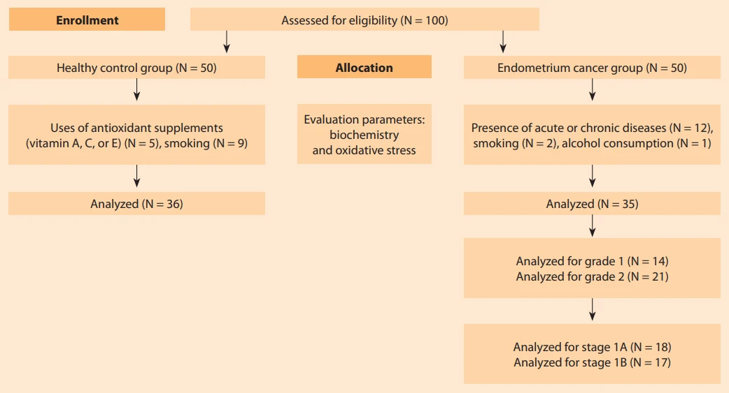
This study was performed at the Necmettin Erbakan University Faculty of Medicine, Department of Obstetrics and Gynecology, between April 15, 2025 and November 15, 2025, using a prospective design. It included 100 patients (N = 50) with EC Study at all stages and a healthy control group (N = 50). The control group consisted of 50 healthy individuals who visited the gynecology outpatient clinic for routine gynecological examinations. Fifteen patients in the EC group and fourteen patients in the control group were excluded from the study based on the exclusion criteria. After applying the predefined exclusion criteria, 35 patients with histopathologically confirmed EC and 36 age-matched healthy controls were included in the final analysis. Biochemical analyses were performed at the Department of Clinical Biochemistry, Yıldırım Beyazıt University, Ankara, Turkey. The research protocol was conducted in accordance with the Helsinki Declaration.
Power analysis (PASS power analysis and sample size system) was used to determine the required sample size for the study and control groups. Based on the study by Demirseren et al., with an effect size of 0.57, a power of 0.80, and a Type I error of 0.05, it was calculated that 50 participants should be included in each group [14].
Demographic data, including age, body mass index (BMI), gravidity, parity, and menopausal status, were collected via clinical interviews. Serum thiol disulfide and oxidative stress parameters were analyzed in all participants. Preoperative evaluation of all EC patients included a comprehensive medical history, physical and pelvic examinations, Pap smear, pelvic ultrasound, chest X-ray, and abdominal magnetic resonance imaging (MRI). Histopathological diagnosis was established via endometrial biopsy [15]. All patients underwent surgery performed by experienced gynecological oncologists at NEÜ University Hospital.
The surgical management included total hysterectomy with bilateral salpingo-oophorectomy, inspection of the peritoneal cavity, collection of peritoneal washings, and lymphadenectomy when indicated. Adjuvant treatment was given to all patients except those with stage IA disease of grade 1 or 2. Histopathological grading and staging were conducted in accordance with the World Health Organization and FIGO criteria.
Exclusion criteria
To minimize potential confounding factors influencing oxidative stress levels, participants with acute or chronic diseases, those using antioxidant supplements (vitamins A, C, or E), and individuals who smoke or consume alcohol were excluded from the study (Fig. 1).
Laboratory analysis
Venous blood samples were obtained in the morning hours (08 : 00–10 : 00) following a 12-hour overnight fast. After collection, serum was separated by centrifugation at 1,500 × g for 10 minutes and promptly stored at −80 °C until analysis. All archived serum samples were thawed and subjected to thiol-disulfide homeostasis analysis. Measurements were performed using a novel, fully automated spectrophotometric method developed by Erel and Neşelioğlu. Disulfide bonds were reduced with sodium borohydride to expose free functional thiol groups. The remaining sodium borohydride was then quenched with formaldehyde to prevent interaction with 5,5’-dithiobis - (2--nitrobenzoic acid) (DTNB). Total thiol concentrations, encompassing both native and reduced thiol groups, were determined following reaction with DTNB. Dynamic disulfide levels were calculated as half the difference between total thiol and native thiol concentrations. The ratios of disulfide to total thiol, disulfide to native thiol, and native thiol to total thiol were expressed as percentages [12].
Ethical aspects
Ethical approval for this study was granted by the Ethics Committee of Necmettin Erbakan University Faculty of Medicine (Approval No: April 2025/217), and the research was carried out in line with the principles of the Declaration of Helsinki. Each participant provided documented written consent for participation prior to being enrolled in the study.
Statistical analysis
The IBM SPSS Statistics (Version 27) software was used to conduct the statistical analyses (IBM, Armonk, NY, USA, 2011). Normality of the variables was determined using the Kolmogorov-Smirnov/Shapiro-Wilk test. The mean ± standard deviation (SD) was used for continuous variables with a normal distribution, while the median (interquartile range – IQR) was used for non-normal variables. Continuous variables having a normal distribution were compared using the independent samples T-test, whereas variables with a non-normal distribution were compared using the Mann-Whitney U-test. For parameters with a normal distribution, a one-way ANOVA test was employed. A Kruskal-Wallis analysis was conducted to examine those whose distribution was not normal. When comparing categorical variables, the Chi-square test or Fisher’s exact test were used. Correlations between the approaches were assessed using Pearson’s or Spearman’s rank correlation coefficients. A P-value under 0.05 was regarded as indicative of a statistically significant result [16,17].
Results
A total of 71 participants were enrolled in the study based on the predefined inclusion criteria. The patient selection process is illustrated in the flow diagram summarised in Fig. 1. Demographic and clinical characteristics of patients with EC and healthy control subjects are detailed in Tab. 1. There were considerable differences between the two groups with in terms of pathological results (endometrioid endometrial carcinoma, 97.1%), the presence of comorbidities; especially hypertension and diabetes mellitus and the use of antidiabetic medications (all P < 0.001).
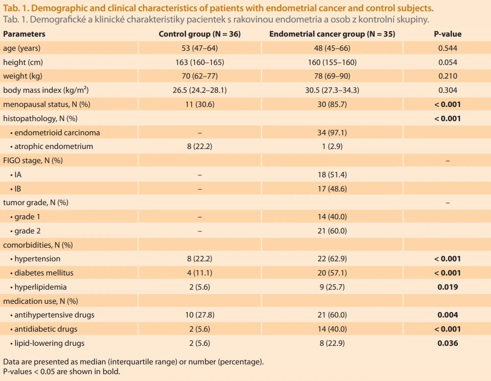
Participants had a median age of 48 years in this study (range, 44–77 years). The most frequently observed parity was two pregnanciesHistopathological evaluation revealed that 60% of patients were diagnosed with Grade 2 disease. Hypertension, diabetes mellitus, and hyperlipidemia were the most common comorbid conditions, with prevalences of 62.9%, 57.1%, and 25.7%, resp. (Tab. 1).
Among patients with EC (N = 35), 18 (51.4%) were classified as stage IA and 17 (48.6%) as stage IB. Of the patients with stage IA disease, 11 had Grade 1 tumors and 7 had Grade 2 tumors. In contrast, the majority of patients with stage IB disease had Grade 2 tumors (15 of 17), while only 2 patients were classified as Grade 1.
Biochemical parameters of the EC and control groups are compared in Tab. 2. In comparison with controls, individuals with EC showed reduced total and native thiol levels (both P < 0.001). Furthermore, HbA1c as well as fasting glucose concentrations were greater in subjects with EC than in healthy individuals (P < 0.001, and P = 0. 004, resp.).
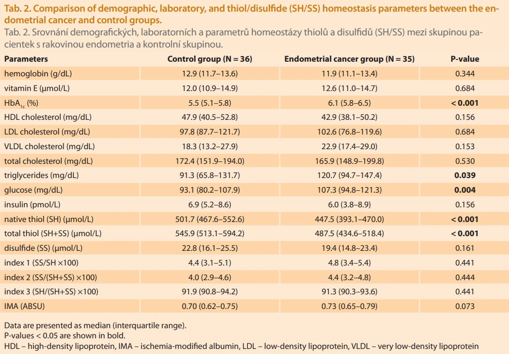
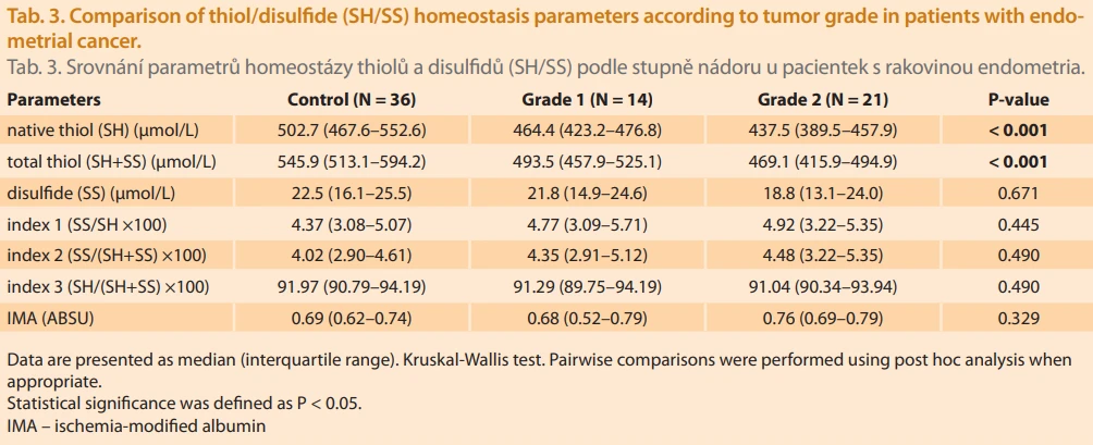
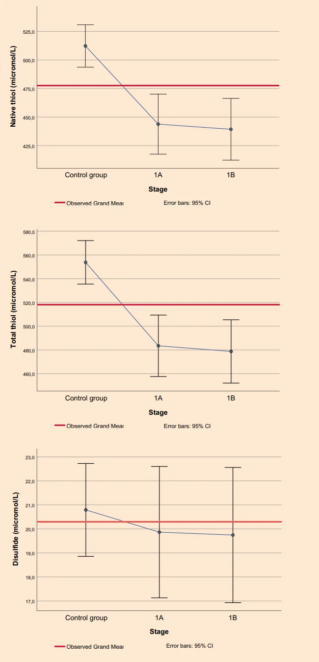
The comparison of thiol/disulfide (SH/SS) homeostasis parameters according to tumor grade is presented in Tab. 3. Patients with Grade 2 EC exhibited significantly lower total and native thiol levels compared with both Grade 1 patients and the control group (P < 0. 001 for all comparisons).
Correlation analysis demonstrated a significant negative association between EC stage and levels of native as well as total thiols (R = −0.586, P < 0.001 and R = −0.604, P < 0.001, resp.). Consistent with this finding, patients with stage IB disease had significantly lower total and native thiol levels compared with the other groups (P < 0.001) (Fig. 2).
Discussions
In the present study, thiol-disulfide homeostasis was investigated in patients with EC and compared with a control group of women who presented for routine gynecological examinations. This study demonstrates a significant disruption in thiol/disulfide homeostasis in patients with endometrial cancer, supporting the role of oxidative stress in disease pathogenesis and progression. Significantly lower levels of both total and native thiols were observed in the EC group compared with healthy controls, indicating a substantial reduction in antioxidant capacity within the tumor microenvironment.
Oxidative stress occurs when there is a disruption in the balance between the generation of ROS and the body’s ability to counteract them through antioxidant defenses, resulting in damage to cellular components. Thiols, which are antioxidants containing sulfhydryl (R-SH) groups, are crucial for neutralizing ROS produced during oxidative stress. Native thiols can donate hydrogen atoms and form disulfide bonds (RSSR) thereby binding excess oxygen and protecting tissues from oxidative injury. Disulfide bonds, in turn, can be reduced back to native thiols, a reversible process referred to as dynamic thiol/disulfide homeostasis [18]. This dynamic balance within proteins is thought to be essential for the regulation of critical cellular processes, including proliferation, differentiation, detoxification, and degeneration [19]. Dynamic thiol-disulfide homeostasis has been evaluated in various gynecological malignancies; however, studies focusing specifically on EC are limited.
In this study, thiol levels appeared to decrease with increasing tumor grade. Patients with grade 2 disease exhibited significantly lower total and native thiol concentrations compared with both grade 1 patients and the control group. These findings suggest that the oxidative stress burden increases with histological aggressiveness. This observation is consistent with evidence indicating that higher tumor grade is associated with enhanced cellular proliferation, inflammation, and enhanced production of ROS, all of which may contribute to accelerated thiol depletion.
Recent studies also support the contribution of ROS to cancer pathogenesis [20]. A primary objective of cancer treatment is to inhibit cancer cell proliferation and to counteract oxidative stress, which triggers the pathophysiological mechanisms responsible for tumor initiation and progression. Therefore, the assessment of oxidative stress levels may be useful in predicting disease severity and clinical course. In various studies evaluating different cancer types, total thiol and native thiol levels have been identified to be lower in patients with prostate, breast, lung, and endometrial cancers compared with control groups [9,21]. In our study, consistent results were obtained with respect to serum concentrations of both total and native thiols. Biochemical analyses revealed that levels of native as well as total thiols were substantially reduced in the EC group relative to the control group, indicating a marked increase in oxidative stress in these patients. Regarding disulphide levels and thiol - disulphide ratios, which are indicators of oxidative stress, there were no discernible variations between the three groups. The grade 1 and grade 2 groups’ lower native and total thiol levels point to a possible involvement of these antioxidants in the etiology of endometrial cancer. Since thiols are key components of the cellular antioxidant defense system, this decrease is believed to result from enhanced reactive oxygen species production combined with a reduction in the body’s antioxidant capacity [11].
A similar pattern is observed in disease stage. Native and total thiol levels were inversely proportional to tumor stage, with significantly lower thiol levels in patients with stage IB disease compared to those with stage IA disease. These results suggest that deeper myometrial invasion is associated with greater oxidative stress, which may reflect increased tumor burden and impaired redox regulation. Overall, these findings suggest that thiol/disulfide homeostasis in EC may reflect not only the presence of the disease but also prognostically important parameters such as tumor grade and stage.
Diabetes and obesity are well-known risk factors for endometrial cancer, and more intensive medical monitoring in these patients may have contributed to lower glycemic parameters. The higher prevalence of hypertension, diabetes, and thyroid disease among patients with EC underscores the role of metabolic and endocrine dysregulation in carcinogenesis and oxidative stress. Such conditions can exacerbate redox imbalance and may be partly responsible for the observed changes in thiol/disulfide homeostasis.
This study has several limitations. There are several limitations to consider in these data. First, the single-center design limits the ability to establish a causal relationship between thiol-disulfide homeostasis and EC progression, and may hinder the generalizability of residual modifiers to broader ruptures. Second, while strict exclusion criteria were applied to minimize oxidative stress contributors, complete exclusion of residual modifiers such as undifferentiated metabolic changes, nutritional changes, or subclinical inflammatory conditions was not possible.
Third, thiol-disulfide homeostasis and oxidative stress were assessed in terms of a single preoperative time frame; therefore, dynamic changes occurring during disease progression or after treat-ment could not be evaluated. Longitudinal measurements provide more robust information on temporal variations between oxidative stress and tumor spread. Fourth, the magnitudes of the data were limited, particularly in subgroup analyses by tumor grade and stage; this may have reduced the power to detect differences, especially in disulfide levels and thiol-disulfide ratios.
Finally, although thiol-disulfide homeostasis is a well known indicator of systemic oxidative stress, tissue-level markers of oxidative stress in the tumor microenvironment have not been evaluated.
Conclusions
The present findings demonstrate that thiol-disulfide homeostasis is significantly disrupted in EC and is associated with tumor grade and stage. Thiol-based redox parameters may represent accessible and complementary biomarkers for evaluating disease severity and oxidative stress status in patients with endometrial cancer. Further prospective studies are warranted to clarify their prognostic value and clinical applicability.
Submitted/Doručeno: 27. 1. 2026
Accepted/Přijato: 1. 2. 2026
Jule Eriç Horasanlı, MD
Department of Obstetrics and Gynecology
Faculty of Medicine
Necmettin Erbakan University
Abdülhamid Han Street No. 3
42080 Selçuklu/Konya
Turkey
juleesen@gmail.com
Zdroje
1. Bray F, Ferlay J, Soerjomataram I et al. Global cancer statistics 2018: GLOBOCAN estimates of incidence and mortality worldwide for 36 cancers in 185 countries. CA Cancer J Clin 2018; 68 (6): 394–424. doi: 10.3322/caac.21492.
2. Sung H, Ferlay J, Siegel RL et al. Global cancer statistics 2020: GLOBOCAN estimates of incidence and mortality worldwide for 36 cancers in 185 countries. CA Cancer J Clin 2021; 71 (3): 209–249. doi: 10.3322/caac.21660.
3. Lee J, Yeo SG, Lee JM et al. The role of reactive oxygen species in the pathogenesis and treatment of endometrial cancer. Front Med (Lausanne) 2025; 12 : 1662794. doi: 10.3389/fmed.2025.1662794.
4. Błachnio-Zabielska AU, Sadowska P, Zdrodowski M et al. The interplay between oxidative stress and sphingolipid metabolism in endometrial cancer. Int J Mol Sci 2024; 25 (19): 10243. doi: 10.3390/ijms251910243.
5. Ziech D, Franco R, Pappa A et al. Reactive oxygen species (ROS) – induced genetic and epigenetic alterations in human carcinogenesis. Mutat Res 2011; 711 (1–2): 167–173. doi: 10.1016/j.mrfmmm.2011.02.015.
6. Sotgia F, Martinez-Outschoorn UE, Lisanti MP. Mitochondrial oxidative stress drives tumor progression and metastasis: should we use antioxidants as a key component of cancer treatment and prevention? BMC Med 2011; 9 : 62. doi: 10.1186/1741-7015-9-62.
7. Prakash M, Shetty MS, Tilak P et al. Total thiols: biomedical importance and their alteration in various disorders. Online J Health Allied Sci 2009; 8 (2): 2.
8. Circu ML, Aw TY. Reactive oxygen species, cellular redox systems, and apoptosis. Free Radic Biol Med 2010; 48 (6): 749–762. doi: 10.1016/j.freeradbiomed.2009.12.022.
9. Eryilmaz MA, Kozanhan B, Solak I et al. Thiol-disulfide homeostasis in breast cancer patients. J Cancer Res Ther 2019; 15 (5): 1062–1066. doi: 10.4103/jcrt.JCRT_553_17.
10. Prabhu A, Sarcar B, Kahali S et al. Cysteine catabolism: a novel metabolic pathway contributing to glioblastoma growth. Cancer Res 2014; 74 (3): 787–796. doi: 10.1158/0008-5472.CAN-13-1423.
11. Sezgin B, Pirincci F, Camuzcuoglu A et al. Assessment of thiol-disulfide balance in early--stage endometrial cancer. J Obstet Gynaecol Res 2020; 46 (7): 1140–1147. doi: 10.1111/jog.14301.
12. Erel O, Neselioglu S. A novel and automated assay for thiol/disulfide homeostasis. Clin Biochem 2014; 47 (18): 326–332. doi: 10.1016/j.clinbiochem.2014.09.026.
13. Wu Y, Wang J, Wang H et al. Study on expression of Ki-67 and early apoptotic protein M30 in endometrial carcinoma and their correlation with prognosis. Zhonghua Bing Li Xue Za Zhi 2003; 32 (4): 314–318.
14. Demirseren DD, Cicek C, Alisik M et al. Dynamic thiol/disulphide homeostasis in patients with basal cell carcinoma. Cutan Ocul Toxicol 2017; 36 (3): 278–282. doi: 10.1080/15569527.2016.1268150.
15. Williams PM, Gaddey HL. Endometrial biopsy: tips and pitfalls. Am Fam Physician 2020; 101 (9): 551–556.
16. Aldrich JO. Using IBM SPSS statistics: an interactive hands-on approach. Thousand Oaks: Sage Publications 2018.
17. de Winter JC, Gosling SD, Potter J. Comparing the Pearson and Spearman correlation coefficients across distributions and sample sizes: a tutorial using simulations and empirical data. Psychol Methods 2016; 21 (3): 273–290. doi: 10.1037/met0000079.
18. Coskun A, Ercan O, Arikan DC et al. Modified Ferriman-Gallwey hirsutism score and androgen levels in Turkish women. Eur J Obstet Gynecol Reprod Biol 2011; 154 (2): 167–171. doi: 10.1016/j.ejogrb.2010.10.001.
19. Beyazit F, Yilmaz N, Balci O et al. Evaluation of oxidative stress in women with polycystic ovary syndrome as represented by serum ischemia-modified albumin and its correlation with testosterone and insulin resistance. Intern Med 2016; 55 (17): 2359–2364. doi: 10.2169/internalmedicine.55.6265.
20. Hizal M, Sendur MA, Aksoy S et al. Evaluation of dynamic serum thiol/disulfide homeostasis in locally advanced and metastatic gastric cancer. J Oncol Sci 2018; 4 (1): 1–4. doi: 10.1016/j.jons.2018.01.002.
21. Sezgin B, Pirincci F, Camuzcuoglu A et al. Thiol-disulfide status of patients with cervical cancer. J Obstet Gynaecol Res 2020; 46 (11): 2423–2429. doi: 10.1111/jog.14480.
Štítky
Dětská gynekologie Gynekologie a porodnictví Reprodukční medicínaČlánek vyšel v časopise
Česká gynekologie
2026 Číslo 4
Nejčtenější v tomto čísle
- Oxidační stres a nerovnováha antioxidantů v endometriální karcinogenezi
- Ektopický zbytek kůry nadledviny ve vejcovodu – vzácný náhodný nález
- Zlepšují mezenchymální kmenové buňky odvozené z tukové tkáně reprodukční potenciál u pacientek s neadekvátní ovariální odpovědí?
- Ultrazvukové vyšetrenie počas pôrodu – prospektívna pilotná komparatívna štúdia presnosti vaginálneho a ultrasonografického vyšetrenia