
Ultrasound examination during labor – a prospective pilot comparative study of clinical and ultrasonographic accuracy
Authors:
L. Borovska 1,2
 ; Jozef Záhumenský 1
; P. Papcun 1; R. Dudič 2
; P. Urdzik 2
; Jozef Záhumenský 1
; P. Papcun 1; R. Dudič 2
; P. Urdzik 2
Authors place of work:
2nd Department of Obstetrics and Gynecology, Faculty of Medicine, Comenius University and University Hospital, Bratislava, Slovakia
1; Department of Obstetrics and Gynecology, University Hospital of L. Pasteur, Košice, Slovakia
2
Published in the journal:
Ceska Gynekol 2026; 91(4): 285-289
Category:
Původní práce
doi:
https://doi.org/10.48095/cccg2026285
Summary
Objective: The aim of this pilot study was to evaluate and compare the diagnostic accuracy of digital vaginal examination versus intrapartum ultrasound (IUS) during the first and second stages of labor in women with cephalic presentation. Materials and methods: This prospective comparative study was conducted at the Department of Gynecology and Obstetrics, L. Pasteur University Hospital in Košice. The cohort included parturients (regardless of parity) with regular uterine contractions, cephalic presentation, and ruptured membranes. To ensure high data quality, all ultrasound examinations were performed by senior obstetricians with at least 8 years of clinical experience. Examining obstetricians were blinded to each other’s findings. The assessment focused on fetal position, head position, head station, and progression during labor. IUS included a transabdominal approach (head position) and a transperineal approach, measuring head-perineum distance and angle of progression. In cases of discrepancy, the ultrasound finding was considered the reference standard. Results: A total of 47 patients were included. Agreement between IUS and VE across all parameters was observed in 27 cases (57%). Discrepancies were noted in 20 patients (43%), with the most frequent errors occurring in the assessment of fetal head position. The statistical superiority of ultrasound over digital examination was highly significant (P < 0.0001). Presence of a caput succedaneum was a major confounding factor, which was present in 45% of non-correlated cases. The caesarean section rate in the cohort was 21% (N = 10). Conclusion: While vaginal examination remains the global gold standard in routine obstetrics, it is inherently subjective and prone to inter-observer variability. Our findings suggest that intrapartum ultrasound provides a more objective, accurate, and reproducible assessment. Incorporating IUS into labor management may reduce the need for frequent digital examinations, potentially lowering the risk of chorioamnionitis and enhancing patient comfort while optimizing clinical decision-making.
Keywords:
labor – intrapartum ultrasound – vaginal examination – labor management – obstetric ultrasound
Introduction
Labor is a dynamic physiological process, the onset of which is often difficult to define, yet its conclusion is marked by the expulsion of the fetus and placenta. While labor is inherently physiological, medical intervention becomes essential in high-risk pregnancies and pathological obstetric conditions. Historically, digital vaginal examination (VE) has been considered the “gold standard” for monitoring labor progress. However, VE is inherently subjective, has limited accuracy, and requires systematic training and extensive clinical experience to achieve proficiency.
Literature reports indicate that the error rate in determining fetal head position via digital examination is between 30% and 50% [1]. In contrast, intrapartum ultrasound (IUS) has emerged as a modern, objective, and reproducible tool that is particularly valuable for less experienced clinicians. By utilizing quantitative parameters, IUS eliminates the subjective nature of fetal head station estimation and facilitates evidence-based labor management. Current clinical indications for IUS, as defined by the International Society of Ultrasound in Obstetrics and Gynecology (ISUOG), include the prolongation of the first or second stage of labor, assessment of malpresentations, evaluation of fetal head station before operative vaginal delivery, and cases complicated by a caput succedaneum.
Beyond its diagnostic utility, ultrasound serves as an effective educational tool for improving manual palpation skills. Furthermore, by reducing the frequency of digital examinations, IUS may decrease the incidence of chorioamnionitis and improve overall patient comfort. Implementing objective data into clinical decision-making has the potential to optimize delivery outcomes and reduce the rate of unnecessary cesarean sections. The aim of our study was to compare the diagnostic accuracy of digital vaginal examination with intrapartum ultrasound and to identify the specific clinical factors contributing to diagnostic discrepancies in the labor ward.
Material and methods
Study design and ethics
This prospective comparative study was conducted at the Department of Gynecology and Obstetrics, L. Pasteur University Hospital in Košice. The study protocol was approved by the Ethics Committee of UNLP, and all participants provided written informed consent prior to enrollment.
Participants
The study included women in labor (regardless of parity) with regular uterine activity during the first and second stages of labor, characterized by cephalic presentation and ruptured membranes. Patients who underwent only one of the examinations were excluded from the analysis.
Clinical and ultrasound assessment
To ensure the reliability of the findings, the examining obstetricians were blinded to the results of the alternative examination. Clinical VE was compared against IUS. To establish IUS as a robust reference standard, all scans were performed by obstetricians with at least 8 years of specialized experience in intrapartum sonography.
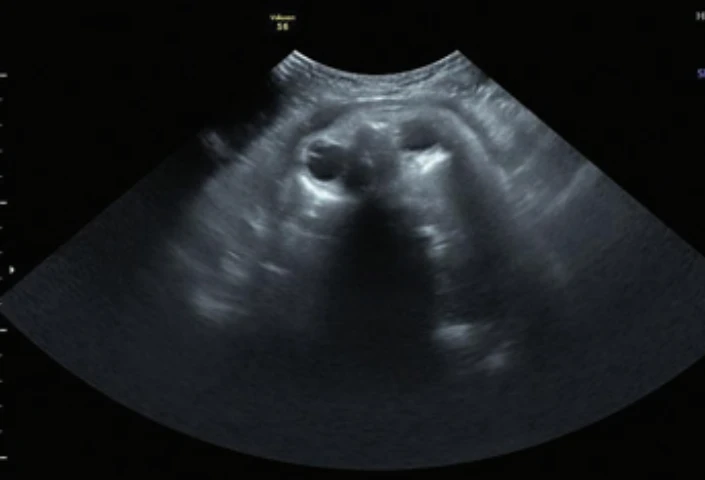
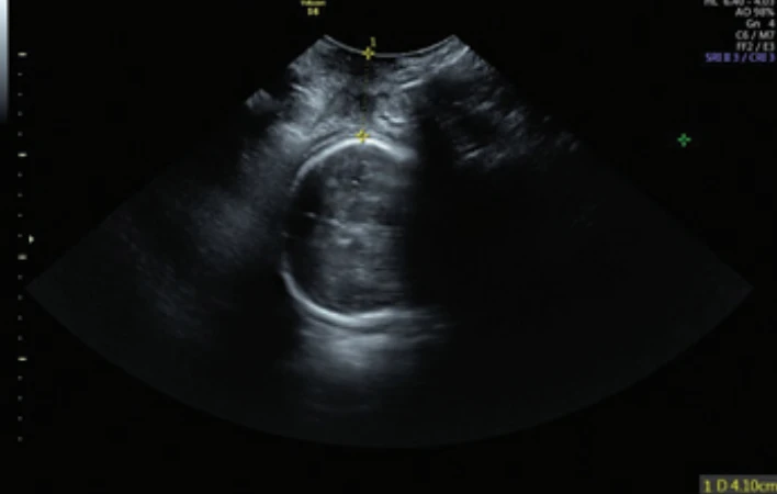
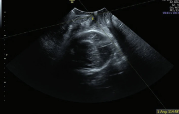
The assessment included fetal position, head station (Fig. 1), and head progression through the birth canal. The ultrasound protocol utilized a transabdominal approach to assess fetal head position and a transperineal approach to measure the head-perineum distance (HPD) (Fig. 2) and the angle of progression (AoP) (Fig. 3).
Data analysis and reference points
In cases of diagnostic discrepancy, the ultrasound finding was considered the reference standard. The fetal position at the time of birth was used for definitive confirmation. While we acknowledge the potential for spontaneous fetal rotation during labor, the high level of agreement between our IUS findings and the position at delivery suggests that intrapartum sonography is a robust predictor of the final delivery position, especially when performed in the late stages of labor.
Equipment
Examinations were performed using GE Voluson S6 and Esaote MyLab 60 XVision devices, both equipped with a convex probe (2–5 MHz).
Results
A total of 47 patients were included in the study. Full agreement between ultrasound and VE across all monitored parameters was observed in 27 cases (57%) (Tab. 1). In the remaining 20 patients (43%), findings were discordant in at least one parameter. The most frequent inaccuracy during digital examination was the determination of fetal head position. Using intrapartum ultrasound as the reference standard, the deviation in VE findings was found to be statistically highly significant (P < 0.0001).
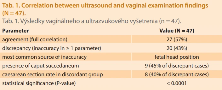
The presence of a caput succedaneum was a major contributor to diagnostic discrepancy, identified in nine patients (45%) within the discordant group. Our data suggest that caput succedaneum significantly increases the risk of incorrect assessment of fetal station and progression, particularly when fontanelles and the sagittal suture are obscured by edema.
The mean maternal age was 25.89 years (range: 16–41 years) (Tab. 2). The cohort comprised 48% primiparas and 52% multiparas. The overall cesarean section (CS) rate was 21% (N = 10). Notably, 80% of these CS cases (8/10) occurred in the group with discordant findings, while only 20% (2/10) occurred in the group with concordant findings.
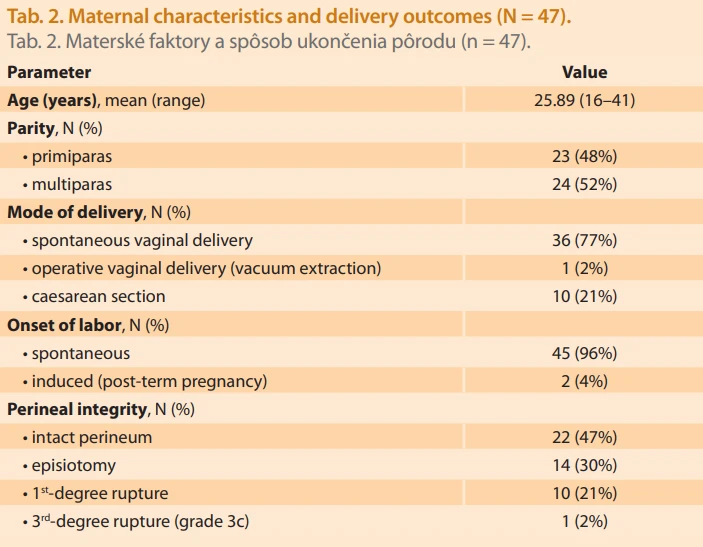
Labor began spontaneously in 45 cases, with two inductions for a post-term pregnancy. Two patients successfully achieved vaginal birth after cesarean (VBAC). One operative vaginal delivery (vacuum extraction) was recorded, complicated by a grade 3c perineal rupture. Regarding overall perineal trauma, 47% of deliveries were without perineal injury, while episiotomy (30%) and first-degree tears (21%) were the most common interventions/injuries. One delivery in a multiparous woman with a hypotrophic fetus was associated with a brow presentation (deflection) and proceeded spontaneously.
At the onset of labor, fetal head positions were distributed as follows: occiput posterior (50%), occiput transverse (37%), and occiput anterior (13%). All cases, excluding one brow presentation that proceeded spontaneously, eventually rotated to the OA position for delivery.
Two deliveries were managed by a midwife, while the remainder were attended by a physician.
Discussion
Digital VE remains a cornerstone of obstetric practice worldwide. Despite its essential role in assessing the progress of labor, it is often stressful and emotionally challenging for mothers, especially when repeated or performed in a rough manner. Evidence suggests that women prefer examinations by female staff or familiar clinicians and prioritize a gentle, expert approach [2]. Furthermore, frequent VE is not only associated with increased pain perception [3] but also poses a clinical risk; performing eight or more examinations during labor has been shown to increase the risk of chorioamnionitis by 1.7-fold [4,5]. The World Health Organization (WHO) therefore recommends limiting the number of vaginal examinations during labor [5]. While our study cohort did not exhibit increased infectious morbidity, the potential to reduce the frequency of invasive examinations through IUS is a significant clinical advantage.
IUS offers an objective and more precise alternative to VE, providing a real-time visualization of the fetal head position, degree of flexion, and descent. This objectivity reduces inter-observer variability and enhances maternal acceptance [3,4,6,7]. Accurate assessment is particularly critical before operative vaginal delivery to avoid complications. In our study, the agreement rate between VE and IUS was 57%, which is lower than that reported by Ramphul et al. [8]. This discrepancy can be attributed to the high prevalence of caput succedaneum in our discordant group (45%). This finding reinforces the notion that soft tissue edema significantly compromises the accuracy of digital palpation, a challenge that IUS effectively overcomes.
Research by Ramphula et al. demonstrated that diagnostic accuracy for head position increases from 79.8% with VE alone to 98.4% when combined with IUS [8]. Incorrect application of forceps or vacuum extractors due to misidentified head position can lead to asynclitism, deflexion, and increased perinatal morbidity [7,9]. In our cohort, the fact that 80% of cesarean sections occurred in cases with discordant findings underscores the potential of IUS to identify stalled labor progress more reliably than VE.
Transperineal ultrasound allows quantitative assessment of parameters such as HPD and AoP. Consistent with the literature, HPD < 40 mm and AoP > 110° are strong predictors of spontaneous vaginal delivery in primiparas [10]. Integrating these measurements into a “sonopartogram” could standardize labor monitoring and reduce subjective error. Additionally, the use of simulators (e. g., IUSIM, LaborPro) provides a valuable educational pathway for clinicians to master these techniques outside the delivery room.
The primary limitation of this study is its relatively small sample size. However, this work was conceived as a pilot study to highlight emerging trends in the labor ward setting. As such, the findings should be interpreted as preliminary, serving as a catalyst for more extensive research with greater statistical power. Despite the small cohort, several key correlations – most notably the impact of caput succedaneum and the discrepancy in CS cases – reached statistical significance, suggesting that the observed effects are strong enough to warrant further investigation.
Conclusion
The rising rate of cesarean sections, frequently indicated due to failure to progress or diagnostic inaccuracies, remains a significant challenge in modern obstetrics. Our study demonstrates that intrapartum ultrasound is a reliable, objective, and reproducible tool that complements – and in complex clinical scenarios, surpasses – traditional vaginal examination. By providing a more precise determination of fetal head position and station, sonography allows for a better prediction of the delivery mechanism and has the potential to reduce the incidence of unnecessary surgical interventions.
The 43% discrepancy between clinical and ultrasound findings identified in this study underscores the role of ultrasound as a critical corrective factor in labor management. While intrapartum ultrasound is not yet universally standardized, its systematic implementation and the continuous education of healthcare professionals are essential. These advancements can lead to optimized labor management, a reduction in cesarean section rates, and an overall improvement in the quality of perinatal care. Further large-scale research is warranted to monitor the long-term impact of this diagnostic modality on perinatal outcomes. Clinical examination remains inherently subjective; therefore, identifying objective methods to enhance safety during labor is essential.
Contribution to authorship
LB, JZ – conceived the study, performed the clinical and ultrasound examinations, analyzed the data, and wrote the main manuscript text.
PP, RD, PU – provided critical revisions and supervised the research. All authors reviewed and approved the final manuscript.
All authors contributed to the study conception and design.
Submitted/Doručené: 13. 2. 2026
Accepted/Prijaté: 16. 2. 2026
prof. Jozef Zahumensky, MD, PhD., MPH
2nd Department of Obstetrics and Gynecology
Faculty of Medicine
Comenius University
University Hospital
Ružinovská 6
821 06 Bratislava
Slovakia
jozef.zahumensky@ru.unb.sk
Zdroje
1. Ghi T, Eggebø T, Lees C et al. ISUOG practice guidelines: intrapartum ultrasound. Ultrasound Obstet Gynecol 2018; 52 (2): 128–139. doi: 10.1002/uog.19072.
2. Lai CH, Levy V. Hong Kong Chinese women’s experiences of vaginal examinations in labour. Midwifery 2002; 18 (4): 296–303. doi: 10.1054/midw.2002.0326.
3. Rizzo G, Aloisio F, Bacigalupi A et al. Women’s compliance with ultrasound in labor: a prospective observational study. J Matern Fetal Neonatal Med 2021; 34 (9): 1454–1458. doi: 10.1080/14767058.2019.1638903.
4. Slagle HB, Hoffman MK, Fonge YN et al. Incremental risk of clinical chorioamnionitis associated with cervical examination. Am J Obstet Gynecol MFM 2022; 4 (1): 100524. doi: 10.1016/j.ajogmf.2021.100524.
5. Hassan WA, Taylor S, Lees C. Intrapartum ultrasound for assessment of cervical dilatation. Am J Obstet Gynecol MFM 2021; 3 (6S): 100448. doi: 10.1016/j.ajogmf.2021.100448.
6. Malvasi A (ed). Intrapartum ultrasonography for labor management. 2nd ed. Switzerland AG: Springer Nature 2021.
7. Eggebø TM, Salvesen KA. Intrapartum sonography and labor progression. In: Malvasi A (ed). Intrapartum ultrasonography for labor management. 2nd ed. Switzerland AG: Springer Nature 2021 : 109–117.
8. Ramphul M, Ooi PV, Burke G et al. Instrumental delivery and ultrasound: a multicentre randomised controlled trial of ultrasound assessment of the fetal head position versus standard care as an approach to prevent morbidity at instrumental delivery. BJOG 2014; 121 (8): 1029–1038. doi: 10.1111/1471-0528.12810.
9. Chan VY, Lau WL. Intrapartum ultrasound and the choice between assisted vaginal and cesarean delivery. Am J Obstet Gynecol MFM 2021; 3 (6S): 100439. doi: 10.1016/j.ajogmf.2021. 100439.
10. Kahrs BH, Eggebø TM. Intrapartum ultrasound in women with prolonged first stage of labor. Am J Obstet Gynecol MFM 2021; 3 (6S): 100427. doi: 10.1016/j.ajogmf.2021.100427.
11. Koterová K, Krofta L, Velebil P et al. Využití ultrazvuku v peripartálním období. Ceska Gynekol 2011; 76 (6): 453–457.
Štítky
Dětská gynekologie Gynekologie a porodnictví Reprodukční medicínaČlánek vyšel v časopise
Česká gynekologie
2026 Číslo 4
Nejčtenější v tomto čísle
- 150 years of the Apolinář maternity hospital in Prague – the history of one of the longest-running maternity hospitals in the world
- Comparative diagnostic accuracy of office hysteroscopy and three-dimensional transvaginal ultrasonography versus histopathology in women with abnormal uterine bleeding
- Personalized approach to infertility treatment in a patient with polyendocrine metabolic ovarian syndrome and chronic pancreatitis
- Ultrasound examination during labor – a prospective pilot comparative study of clinical and ultrasonographic accuracy