
Ectopic adrenal cortical rest in the fallopian tube – a rare incidental finding
Authors:
A. Karabulut
 ; D. Sezen
; A. H. İnan
; D. Sezen
; A. H. İnan
Authors place of work:
Department of Obstetrics and Gynecology, University of Health Sciences Tepecik Training and Research Hospital, Izmir, Turkey
Published in the journal:
Ceska Gynekol 2026; 91(4): 326-328
Category:
Kazuistika
doi:
https://doi.org/10.48095/cccg2026326
Summary
Ectopic adrenal tissue (EAT) is a rare developmental anomaly most frequently observed along the genitourinary tract, usually in pediatric males. Its persistence into adulthood, particularly within the fallopian tube, is exceedingly uncommon. We present the case of a 43-year--old woman who underwent total laparoscopic hysterectomy and bilateral salpingectomy for symptomatic uterine leiomyomas. Histopathologic examination incidentally revealed a well-circumscribed nodule in the right fallopian tube composed of polygonal cells with clear cytoplasm, morphologically resembling adrenal cortical tissue. Immunohistochemical staining demonstrated strong positivity for inhibin and calretinin, confirming the diagnosis of an ectopic adrenal cortical rest. No clinical or biochemical evidence of hormonal hyperfunction was noted. This rare finding expands the limited body of literature on fallopian tube EAT, emphasizing the importance of recognizing this entity to avoid misdiagnosis with metastatic clear cell lesions.
Keywords:
fallopian tube – ectopic adrenal tissue – adrenal cortical rest
Introduction
Ectopic adrenal tissue (EAT), also known as adrenal cortical rest, refers to the presence of adrenocortical cells outside the normal adrenal gland. First described by Morgagni in 1740 [1], EAT represents a developmental anomaly caused by aberrant migration or separation of adrenocortical primordial cells during embryogenesis [2,3]. It occurs in approximately 50% of neonates but typically regresses within the first year of life [3,4]. Persistence into adulthood is rare, with an incidence of around 1% [5]. EAT is most frequently found along the genitourinary tract, including the kidneys, retroperitoneum, broad ligament, spermatic cord, and testes [2,3,6]. In females, parasalpingeal or paraovarian locations have occasionally been described, but true localization within the fallopian tube wall remains extremely uncommon [6–8]. Only a handful of such cases have been reported to date [3,4,8]. Although most lesions are asymptomatic and found incidentally, EAT may rarely undergo hyperplasia or neoplastic transformation, resulting in hormone production or tumor formation [9]. Because its morphology may mimic metastatic clear cell carcinoma or ovarian hilus cell heterotopia, recognizing this entity is essential to prevent misdiagnosis [6–8]. Here, we report a rare case of ectopic adrenal cortical tissue identified incidentally within the fallopian tube of an adult woman and review the relevant literature.
Case report
A 43-year-old woman presented to our gynecology clinic with abnormal uterine bleeding and chronic pelvic pain persisting for several months. Her obstetric history included one cesarean section, prior ovarian cystectomy, and myomectomy. She had no history of endocrine disorders or medication use. On clinical examination, the abdomen was soft and non-tender, with no palpable adnexal masses. Transvaginal ultrasonography revealed an enlarged uterus containing multiple intramural and subserosal leiomyomas, while both ovaries appeared normal. Routine hematologic and biochemical parameters, including serum electrolytes and hormonal assays, were within normal limits.
After counseling, the patient underwent total laparoscopic hysterectomy with bilateral salpingectomy for symptomatic uterine fibroids unresponsive to medical management. The procedure was performed under general anesthesia using a standard four-port approach. Intraoperatively, the uterus was bulky and nodular, consistent with multiple leiomyomas. Both adnexa appeared normal, and no peritoneal or pelvic abnormalities were observed. The postoperative course was uneventful, and the patient was discharged on the second postoperative day.
Histopathologic examination revealed multiple leiomyomas and foci of adenomyosis within the uterine wall. Incidentally, a well-circumscribed, yellowish nodule measuring 2 mm was identified in the right fallopian tube. Microscopically, the nodule consisted of nests and cords of polygonal cells with abundant clear to amphophilic cytoplasm, central round nuclei, and fine granular chromatin, morphologically resembling adrenal cortical cells (Fig. 1–3). No mitoses, cytologic atypia, or necrosis were observed. Immunohistochemical staining showed diffuse positivity for inhibin and calretinin, while cytokeratin and synaptophysin were negative, confirming the diagnosis of ectopic adrenal cortical rest.
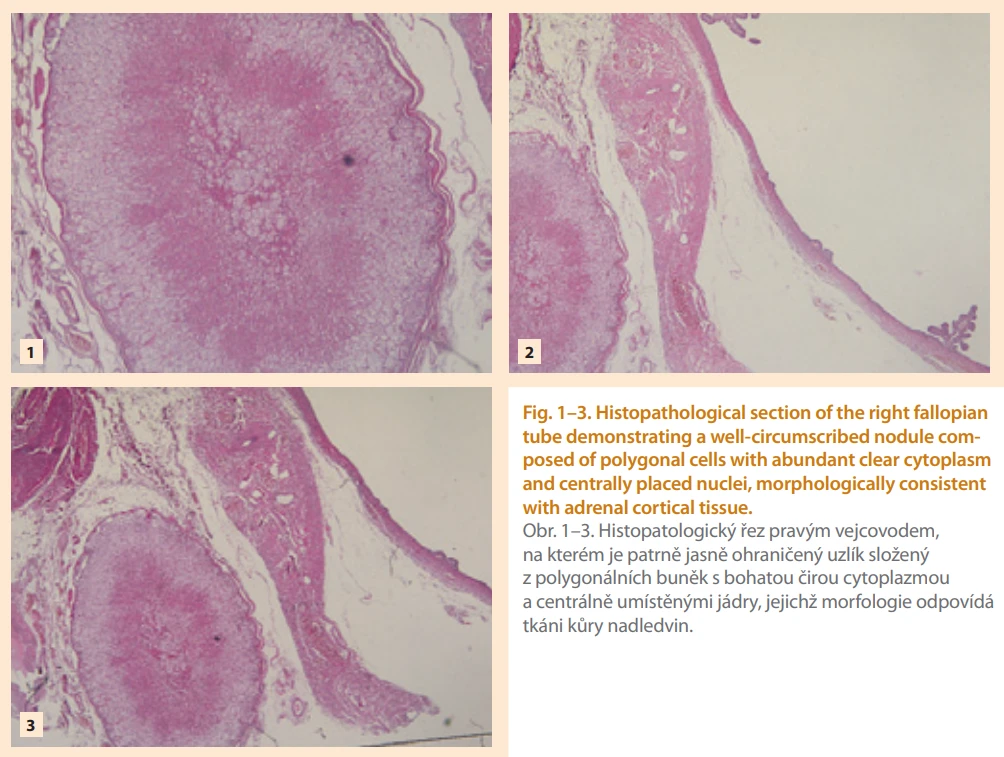
Postoperatively, the patient was clinically stable and showed no evidence of abnormal hormonal secretion. She remained asymptomatic during a six--month follow-up period with no recurrence or endocrine manifestations.
Discussion
Ectopic adrenal cortical rests originate from aberrant migration or incomplete separation of adrenocortical primordial cells during embryogenesis [1–3]. Because of their proximity to the developing gonadal ridge, small cortical fragments can become sequestered along the path of gonadal descent and persist as EAT. These rests are most often seen in the kidneys, broad ligament, and spermatic cord but are exceedingly rare in the fallopian tube [3,4,8].
Although EAT occurs in up to half of neonates, it usually regresses in infancy [3,4]. Persistence into adulthood is rare, with a prevalence of approximately 0.07% in adult urogenital surgical specimens [2]. Reported fallopian tube cases, such as those by Tingi and Ogah (2018), Tzigkalidis et al. (2021), Hafiz and Alturkistani (2022), Al-Janabi et al. (2024), and Gaffoor et al. (2025), were all incidental findings during surgery for benign conditions including leiomyoma, adenomyosis, or ectopic pregnancy [6–8].
Histologically, EAT presents as well-circumscribed nodules of polygonal cells with clear or finely granular cytoplasm resembling adrenal cortical tissue [6,9]. Immunohistochemistry is critical for confirmation, as it differentiates EAT from metastatic renal cell carcinoma and ovarian hilus cell heterotopia. Positivity for inhibin, calretinin, and Melan-A, and negativity for cytokeratin and synaptophysin, confirm its adrenocortical origin [9,10]. Our findings of inhibin and calretinin positivity without atypia or mitosis are consistent with previously reported features.
Although most cases are nonfunctional, ectopic adrenal rests may rarely undergo hyperplasia or neoplastic transformation under chronic ACTH stimulation or hormonal imbalance [9]. Functional transformation has been described mainly in pediatric or male patients, but no hormonally active lesions have been documented in the fallopian tube. Our patient remained asymptomatic throughout follow-up.
Recognition of this lesion is important to avoid misinterpretation, as its morphology may mimic metastatic clear cell carcinoma. Awareness of this benign entity among gynecologic surgeons and pathologists can prevent unnecessary investigations and overtreatment [6–8]. This case contributes to the limited literature on fallopian tube EAT and underscores the importance of meticulous histopathologic examination of adnexal specimens – even when grossly unremarkable – to detect rare but educational developmental anomalies.
Conclusion
Ectopic adrenal cortical tissue in the fallopian tube is an exceedingly rare incidental finding in adult women. Recognition of this lesion is essential, as it can mimic metastatic clear cell carcinoma or other clear cell proliferations. Immunohistochemical analysis with markers such as inhibin, calretinin, and Melan-A is vital for accurate diagnosis. Although most cases are clinically silent, awareness of this entity remains important because ectopic adrenal rests may occasionally undergo hyperplasia or neoplastic transformation [9]. Our case adds to the very limited number of reports describing this anomaly and emphasizes the need for careful histopathologic evaluation of all adnexal tissues. Greater awareness among clinicians and pathologists will help prevent misdiagnosis and improve understanding of ectopic adrenal pathology in the female genital tract.
Submitted/Doručeno: 25. 12. 2025
Accepted/Přijato: 1. 2. 2026
Alaattin Karabulut, MD
Department of Obstetrics and Gynecology
University of Health Sciences
Tepecik Training and Research Hospital
Izmir, 35100
Turkey
alaattin_karabulut@hotmail.com
Zdroje
1. Anderson JR, Ross AH. Ectopic adrenal tissue in adults. Postgrad Med J 1980; 56 (661): 806–808. doi: 10.1136/pgmj.56.661.806.
2. Falco EC, Daniele L, Metovic J et al. Adrenal rests in the uro-genital tract of an adult population. Endocr Pathol 2021; 32 (3): 375–384. doi: 10.1007/s12022-021-09685-y.
3. Tzigkalidis T, Skandalou E, Manthou ME et al. Adrenal cortical rests in the fallopian tube: report of a case and review of the literature. Medicines (Basel) 2021; 8 (3): 14. doi: 10.3390/medicines8030014.
4. Al-Janabi MH, Salamah D, Suleiman M et al. Ectopic adrenal tissue in the mesosalpinx of an older female: the fourth case report in the literature. Oxf Med Case Reports 2024; 2024 (4): omae024. doi: 10.1093/omcr/omae024.
5. Chakrabarti I, Das N. Ectopic adrenal rest in paratubal region: a rare lesion in a rare location. J Microsc Ultrastruct 2022; 11 (2): 118–120. doi: 10.4103/jmau.jmau_139_20.
6. Hafiz B, Alturkistani F. Adrenal cortical rests in the fallopian tube: a case report and review of the literature. Cureus 2022; 14 (8): e27649. doi: 10.7759/cureus.27649.
7. Gaffoor N, Shetty A, Vidhya V et al. Bilateral adrenal cortical rests: an interesting innocuous intruder in the fallopian tubes. Indian J Pathol Microbiol 2025; 68 (2): 402–404. doi: 10.4103/ijpm.ijpm_976_23.
8. Tingi E, Ogah J. Ectopic adrenal rest cells of the fallopian tube: a case report and review of the literature. J Obstet Gynaecol 2018; 38 (4): 578–579. doi: 10.1080/01443615.2017.1379063.
9. Kasajima A, Nakamura Y, Adachi Y et al. Oncocytic adrenocortical neoplasm arising from adrenal rest in the broad ligament of the uterus. Pathol Int 2014; 64 (4): 183–188. doi: 10.1111/pin.12154.
10. Khandakar B, Dey S, Ray PS et al. Ectopic paratubal adrenal cell rest associated with mucinous cystadenoma of ovary. J Clin Diagn Res 2015; 9 (10): ED13–ED14. doi: 10.7860/JCDR/2015/15411.6638.
Štítky
Dětská gynekologie Gynekologie a porodnictví Reprodukční medicínaČlánek vyšel v časopise
Česká gynekologie
2026 Číslo 4
Nejčtenější v tomto čísle
- Oxidative stress and antioxidant imbalance in endometrial carcinogenesis
- Ectopic adrenal cortical rest in the fallopian tube – a rare incidental finding
- Do adipose-derived mesenchymal stem cells improve reproductive potential in patientswith inadequate ovarian response?
- Ultrasound examination during labor – a prospective pilot comparative study of clinical and ultrasonographic accuracy