Comparative diagnostic accuracy of office hysteroscopy and three-dimensional transvaginal ultrasonography versus histopathology in women with abnormal uterine bleeding
Authors:
M. Fouad 1; M. El Mahy 1; S. K. Hamada 1; M. Ibrahim 1
 ; Y. A. Bayoumi 1; A. Sayed 1; H. M. Hawas 1; A. N. Shaker 2
; N. H. Eisa 1; R. M. Elnaggar 3
; A. A. Z. El-Nizamy - 4
; Y. A. Bayoumi 1; A. Sayed 1; H. M. Hawas 1; A. N. Shaker 2
; N. H. Eisa 1; R. M. Elnaggar 3
; A. A. Z. El-Nizamy - 4
Authors place of work:
Obstetrics and Gynecology Department, Kasr Alainy Hospital, Cairo University, Cairo, Egypt
1; Obstetrics and Gynecology Department, Faculty of Medicine, Kafr El-Sheikh University, Kafr El-Sheikh, Egypt
2; Obstetrics and Gynecology Department, Faculty of Medicine, Tanta University, Tanta, Egypt
3; Obstetrics and Gynecology Department, Faculty of Medicine, Suez University, Suez, Egypt
4
Published in the journal:
Ceska Gynekol 2026; 91(4): 273-284
Category:
Původní práce
doi:
https://doi.org/10.48095/cccg2026273
Summary
Background and aim: Abnormal uterine bleeding (AUB) is a common gynecological complaint with significant clinical and quality--of-life implications. Accurate diagnosis of the underlying pathology is critical to guide management. While three-dimensional transvaginal ultrasonography (3D TVUS) offers non-invasive evaluation of the uterine cavity and myometrium, hysteroscopy remains the gold standard for direct visualization. This study evaluated the diagnostic performance of 3D TVUS and hysteroscopy in women presenting with abnormal uterine bleeding, using histopathology as the reference standard. Methods: This prospective observational cross-sectional study included 80 women presenting with AUB at Cairo University Hospital between June 2021 and April 2023. All participants underwent 3D TVUS, office hysteroscopy, and histopathological evaluation where applicable. Formal comparative analysis with hysteroscopy was restricted to intracavitary lesions, while myometrial lesions were evaluated primarily by TVUS. Diagnostic performance metrics (sensitivity, specificity, PPV – positive predictive value, NPV – negative predictive value, and accuracy) were calculated for individual lesions, overall pathology versus normal, and endometrial cavity lesions. Results: The mean age of participants was 38.6 ± 9.8 years; menorrhagia (30%) was the most common presentation. Endometrial polyps (20%), adenomyosis (17.5%), and intramural fibroids (15%) were the leading pathologies. TVUS showed superior performance for myometrial lesions, with the highest accuracy for intramural fibroids (92.5%). Hysteroscopy was more accurate for intracavitary lesions, particularly hyperplasia (97.5%) and intrauterine adhesions (98.8%). For overall pathology versus normal, TVUS demonstrated higher sensitivity (97.1%) and accuracy (93.8%), while hysteroscopy offered greater specificity (80.0%) but lower NPV (40.0%). Conclusion: 3D TVUS and office hysteroscopy are complementary diagnostic modalities in AUB. TVUS is superior for myometrial lesions, while hysteroscopy remains more accurate for intracavitary pathology. A combined approach offers the most comprehensive diagnostic strategy, optimizing accuracy and minimizing unnecessary invasive procedures.
Keywords:
abnormal uterine bleeding – three-dimensional transvaginal ultrasonography – office hysteroscopy – endometrial pathology – diagnostic accuracy
Introduction
Abnormal uterine bleeding (AUB) is one of the most common gynecological complaints, affecting up to one-third of women during their lifetime, with peaks of incidence at menarche and perimenopause [1]. It is defined as any variation in menstrual cycle regularity, frequency, duration, or volume, occurring outside of pregnancy [1]. According to the International Federation of Gynecology and Obstetrics (FIGO) classification system (PALM-COEIN), AUB etiologies are categorized into structural causes – such as polyps, adenomyosis, leiomyomas, and malignancy/hyperplasia – and nonstructural causes, including coagulopathy, ovulatory dysfunction, endometrial disorders, iatrogenic factors, and not otherwise classified conditions [2]. The condition can adversely affect physical, psychological, social, and sexual quality of life, and is a major indication for gynecological consultations, investigations, and surgical interventions, including hysterectomy [3].
Accurate diagnosis of the underlying cause of AUB is essential to guide appropriate management and avoid unnecessary interventions. Conventional diagnostic modalities include transvaginal ultrasonography (TVUS), hysteroscopy, sonohysterography, and endometrial sampling [4]. While two-dimensional TVUS is widely used as a first-line, noninvasive imaging tool, its sensitivity in detecting focal intrauterine lesions such as polyps or submucosal fibroids is limited [5]. The introduction of three-dimensional transvaginal ultrasonography (3D-TVUS) has enhanced diagnostic capability by providing coronal plane imaging, improved spatial orientation, and volumetric assessment, allowing better visualization of the endometrial cavity and its relationship to the surrounding myometrium [6].
Hysteroscopy, on the other hand, remains the gold standard for direct visualization of the uterine cavity, enabling targeted biopsies and simultaneous therapeutic interventions [7]. Office hysteroscopy offers high diagnostic accuracy for focal lesions and avoids the limitations of blind sampling methods, such as dilatation and curettage. Histopathological examination of biopsy specimens continues to serve as the reference standard where applicable [1]. Given the need for precise, cost-effective, and minimally invasive diagnostic strategies, comparative evaluation of advanced imaging modalities against hysteroscopy and histopathology is of clinical importance.
The present study aimed to evaluate the diagnostic performance of office hysteroscopy and three-dimensional transvaginal ultrasonography in detecting uterine pathology in women presenting with abnormal uterine bleeding, using histopathology as the reference standard where applicable, while additionally highlighting the complementary role of 3D TVUS in detecting myometrial pathologies beyond the diagnostic scope of hysteroscopy.
Patients and methods
This prospective observational cross--sectional study was conducted at the Department of Obstetrics and Gynecology, Cairo University Hospital, over a 22-month period from June 2021 to April 2023. A total of 80 women presenting with abnormal uterine bleeding (AUB) were enrolled. Ethical approval was obtained from the institutional review board (approval number: MD-237-2021), and written informed consent was obtained from all participants prior to inclusion. Eligible participants included women presenting with AUB. Exclusion criteria were hemodynamic instability, severe chronic anemia secondary to prolonged bleeding, known coagulation or bleeding disorders, the presence of cervical lesions or cervical cancer, and pregnancy. All women were admitted for standardized clinical evaluation and diagnostic workup (Fig. 1).

Each participant underwent detailed history taking, including menstrual history, bleeding pattern, menopausal status, and previous gynecological surgery, in addition to physical examination including general, abdominal, and pelvic (bimanual and speculum) examinations, to identify abnormal findings and exclude local causes of bleeding. Laboratory investigations included complete blood count (CBC), coagulation profile, random blood sugar (RBS), and liver and kidney function tests.
All patients initially underwent two--dimensional transvaginal ultrasonography (2D TVS) in the outpatient setting using a General Electric (GE) Logic 200 ultrasound machine equipped with a 6.5-MHz transvaginal probe. Uterine size and endometrial thickness (ET) were measured in the midline sagittal plane at the point of maximal thickness. The endometrial stripe was assessed for contour and classified as normal or abnormal, with documentation of focal lesions such as endometrial polyps, submucous or intramural fibroids, adenomyosis, or thickened endometrium (> 14 mm).
Subsequently, three-dimensional transvaginal ultrasonography (3D TVS) was performed using a Voluson E6 system (USA) equipped with an S-VDW 5–8 MHz transvaginal probe. Following completion of the 2D scan, 3D volumes were acquired through an automated 360° sweep, generating truncated cone-shaped volumes (depth 4.3–8.6 cm, alpha angle 90°). Multiplanar reconstruction allowed simultaneous visualization of axial, sagittal, and coronal planes, facilitating precise localization of focal lesions in relation to the endometrium and surrounding myometrium. Findings were compared with 2D TVS results to confirm detected abnormalities or identify additional lesions.
Office hysteroscopy was performed during the early proliferative phase of the menstrual cycle (day 6–10) using a rigid continuous-flow panoramic hysteroscope (Karl Storz, Germany) with a 5 - mm outer sheath, a 30° telescope, and a 150-W metal halide light source (Circon Acmi G71A, Germany). Saline or glycine was used as the distension medium under manometric control at 100–120 mmHg. All patients received intramuscular diclofenac sodium (Voltaren® 75 mg) 30–60 minutes prior to the procedure for analgesia. Using the vaginoscopic approach, the uterine cavity was systematically examined, and intracavitary abnormalities – including endometrial polyps, submucous fibroids, adhesions, and areas of abnormal endometrial thickening – were documented.
In selected inpatient cases, diagnostic hysteroscopy was performed under general anesthesia by a single experienced operator blinded to ultrasound findings. A rigid panoramic hysteroscope (Endomed Systems) with a 6-mm sheath and saline distension at 150 mmHg was used. The uterine cavity was inspected in a standardized sequence, including the fundus, anterior and posterior walls, lateral walls, and uterotubal junctions. Endometrial features such as thickness, color, vascular pattern, and lesion size and location were recorded.
Histopathological examination of endometrial sampling and/or targeted biopsy served as the reference standard for intracavitary lesions. For myometrial lesions (intramural fibroids and adenomyosis), histopathology was considered the reference standard where tissue confirmation was clinically available/applicable; these lesions were assessed primarily as TVUS-based findings rather than within the formal hysteroscopy comparative framework. The procedure included initial endocervical sampling followed by cervical dilatation (Hegar size 7–8) and fractional curettage using a sharp curette to obtain tissue from the fundus and all uterine walls. Specimens were immediately fixed in 10% formalin and examined by a single experienced pathologist. In cases where focal lesions were identified hysteroscopically, targeted biopsies were obtained using hysteroscopic biopsy forceps or curettes to ensure representative sampling, including the superficial myometrial interface when clinically indicated.
For methodological clarity, hysteroscopy was evaluated exclusively for intracavitary abnormalities, namely endometrial polyps, submucous fibroids, endometrial hyperplasia, and intrauterine adhesions. Intramural fibroids and adenomyosis were assessed solely by TVUS, with histopathology serving as the reference standard where applicable. Intramural fibroids and adenomyosis were not included within the formal comparative diagnostic accuracy framework for hysteroscopy, given its inherent limitation to visualization of the endometrial cavity. Histopathological findings were ultimately used to determine the diagnostic accuracy of 2D TVS, 3D TVS, and office hysteroscopy in women presenting with AUB.
Sample size
Sample size was estimated using Buderer’s formula (1996) for precision-based estimation of sensitivity. We planned to estimate the sensitivity of 3D TVUS for detecting clinically significant endometrial pathology in women with AUB (target condition defined a priori as premalignant/malignant or clinically significant endometrial pathology requiring histopathological confirmation). Based on prior literature by van den Boschab et al. and Shiva et al. [8,9], the expected sensitivity was assumed to be 0.76 (range 0.54–0.982). We selected a two-sided 95% confidence level (Z = 1.96) and a predefined absolute precision (half--width) of L = 0.19 for sensitivity. The anticipated prevalence of the target condition in referred AUB cases was assumed to be approximately 25%. Using Buderer’s equation for sensitivity, the minimum required sample size was approximately 78, and therefore 80 women were enrolled. Calculations were done using Buderer equation (1996) [10].
Statistical analysis
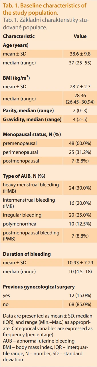
Data were analyzed using descriptive and diagnostic accuracy measures. Continuous variables, including age, body mass index (BMI), parity, gravidity, and duration of bleeding, were assessed for distribution and are presented as mean ± standard deviation (SD) for normally distributed data and as median with range where appropriate. Categorical variables, such as menopausal status, bleeding patterns, and distribution of uterine pathologies, are presented as frequencies and percentages (Tab. 1).
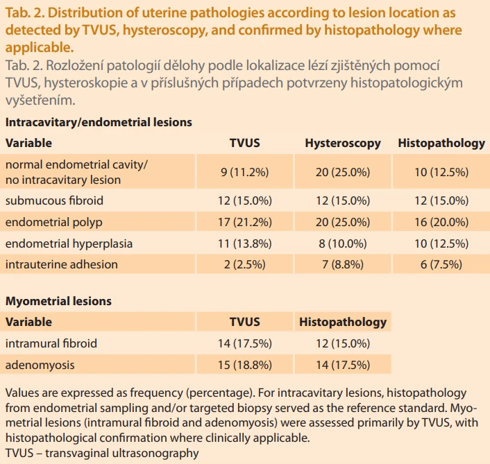
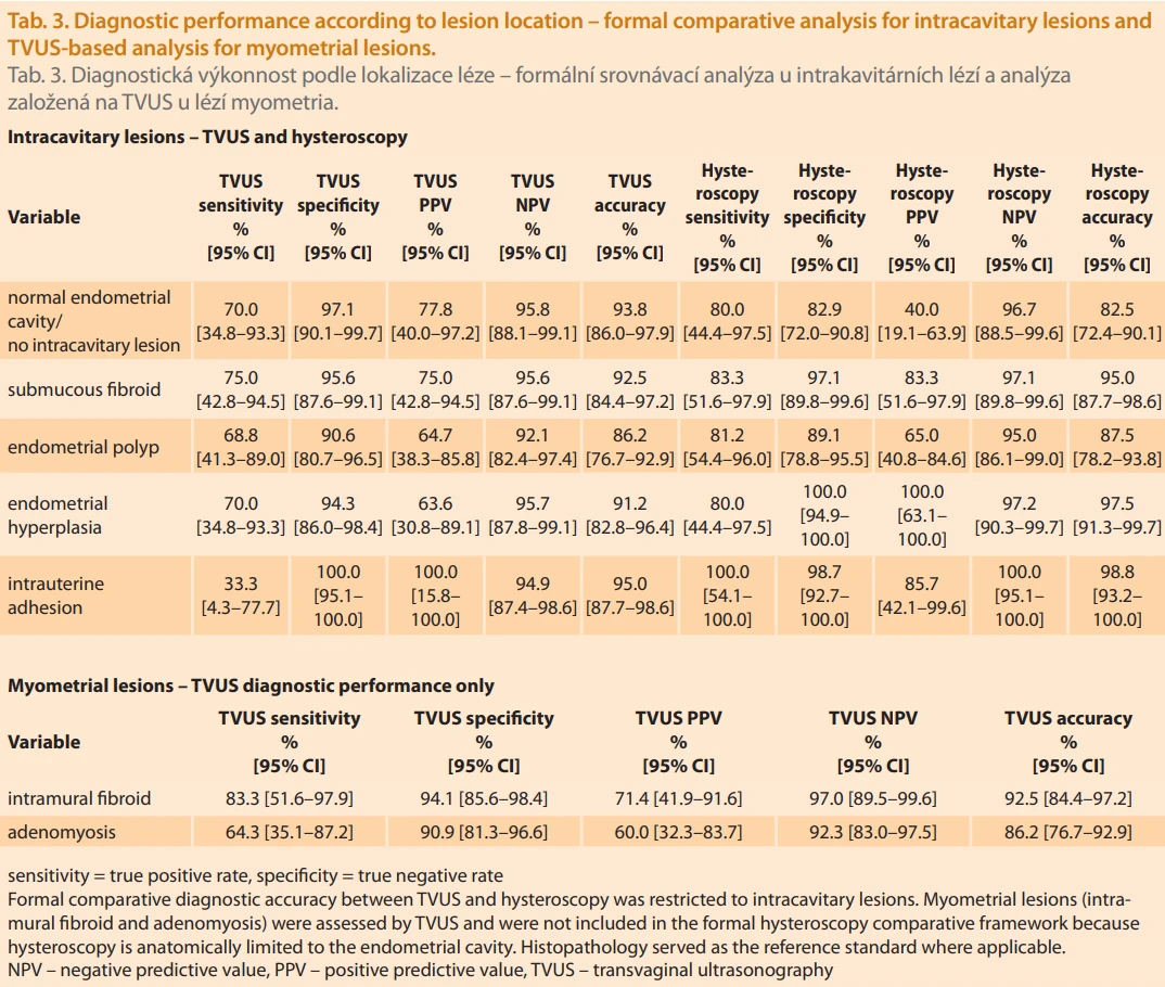
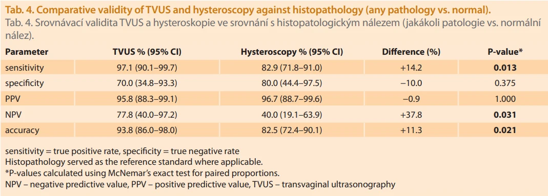
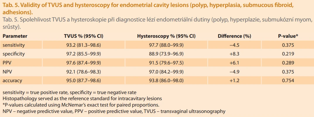
The diagnostic performance of three--dimensional transvaginal ultrasonography (TVUS) and office hysteroscopy was evaluated using histopathological examination as the reference standard for intracavitary lesions and for myometrial lesions where tissue confirmation was clinically available/applicable. For intracavitary lesions, sensitivity, specificity, positive predictive value (PPV), negative predictive value (NPV), and overall accuracy were calculated for both TVUS and hysteroscopy (Tab. 2, 3). For myometrial lesions (intramural fibroids and adenomyosis), diagnostic performance was calculated for TVUS only, as these lesions are outside the anatomical scope of hysteroscopy. In addition, overall pathology versus normal findings was analyzed in Tab. 4, and endometrial cavity lesions were analyzed in Tab. 5. All diagnostic estimates are reported with 95% confidence intervals (CIs), calculated using exact binomial (Clopper-Pearson) methods.
Because both TVUS and hysteroscopy were performed in the same patients, paired comparisons of diagnostic performance were conducted. McNemar’s exact test was used to compare sensitivity, specificity, negative predictive value, and overall accuracy between the two modalities. Differences in diagnostic proportions are presented. A two-sided P-value < 0.05 was considered statistically significant.
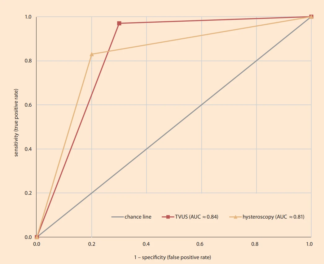
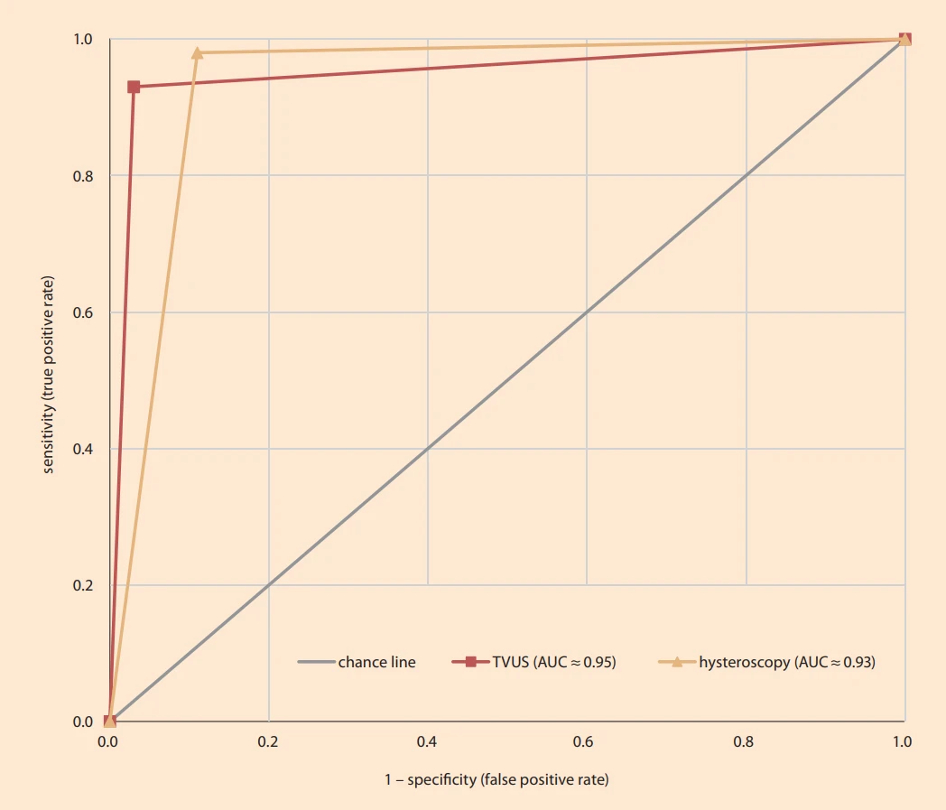
Receiver operating characteristic (ROC) curve analysis was performed to illustrate the trade-off between sensitivity and specificity for each diagnostic modality, and the area under the ROC curve (AUC) was calculated as a summary measure of overall diagnostic performance (Graphs 1, 2). Statistical analyses were conducted using standard statistical software.
Results
Table 1 summarizes the baseline characteristics of the study population. The mean age of participants was 38.6 ± 9.8 years (range: 25–55 years), with many women being premenopausal (60%). The mean body mass index (BMI) was 28.7 ± 2.7 kg/m², indicating that most participants were overweight. The median parity was 2, and the median gravidity was 4. A history of previous gynecological surgery was present in 12 women (15%). The mean duration of bleeding was 10.9 ± 7.3 days, exceeding the normal menstrual duration. Menorrhagia was the most frequent bleeding pattern (30%), followed by metrorrhagia (20%). Postmenopausal women constituted 8.8% of the cohort, a subgroup of clinical importance due to the increased risk of endometrial pathology.
Table 2 presents the distribution of uterine pathologies according to lesion location. Intracavitary lesions are shown in direct comparison between TVUS, hysteroscopy, and histopathology, whereas myometrial lesions are presented as TVUS-assessed findings with histopathological correlation where applicable. Endometrial polyps were the most common lesion (20%), followed by adenomyosis (17.5%) and intramural fibroids (15%). A clear distinction was observed between endometrial cavity lesions and myometrial lesions. TVUS demonstrated higher detection rates for myometrial pathologies, particularly intramural fibroids and adenomyosis, whereas hysteroscopy more frequently identified intrauterine adhesions. Submucous fibroids were detected at comparable rates by both modalities, and endometrial polyps showed similar detection frequencies.
Table 3 summarizes diagnostic performance according to lesion location, with formal comparative analysis between TVUS and hysteroscopy limited to intracavitary lesions, while myometrial lesions were analyzed as TVUS-based findings only. For myometrial lesions, TVUS showed excellent diagnostic performance, achieving the highest accuracy for intramural fibroids (92.5%) and good accuracy for adenomyosis (86.2%). These lesions were assessed as TVUS--detected myometrial pathologies and were not included in the formal comparative diagnostic accuracy framework for hysteroscopy because they lie outside the direct anatomical scope of the endometrial cavity. For endometrial cavity lesions, hysteroscopy demonstrated excellent diagnostic performance, with high accuracy for endometrial hyperplasia (97.5%), submucous fibroids (95.0%), and intrauterine adhesions (98.8%). TVUS also showed strong performance for intracavitary abnormalities but had a notable limitation in detecting intrauterine adhesions, with a sensitivity of 33.3%.
Table 4 compares the overall diagnostic validity of TVUS and hysteroscopy in distinguishing any uterine pathology from normal findings. TVUS demonstrated significantly higher sensitivity (97.1 vs. 82.9%; P = 0.013), significantly higher negative predictive value (77.8 vs. 40.0%; P = 0.031), and significantly higher overall accuracy (93.8 vs. 82.5%; P = 0.021) compared with hysteroscopy.
Although hysteroscopy showed a higher specificity (80.0 % vs 70.0%) and slightly higher PPV (96.7 vs. 95.8%), these differences were not statistically significant (P = 0.375 for specificity, and P = 1.000 for PPV). These findings indicate that TVUS is more reliable for ruling out pathology, while hysteroscopy maintains high confirmatory value when pathology is detected.
Table 5 presents a formal paired comparison of TVUS and hysteroscopy for endometrial cavity lesions (endometrial polyps, hyperplasia, submucous fibroids, and intrauterine adhesions). Both modalities demonstrated high diagnostic validity, with no statistically significant differences between them across all diagnostic parameters.
TVUS showed higher specificity (97.2 vs. 88.9%) and PPV (97.6 vs. 91.5%), whereas hysteroscopy achieved slightly higher sensitivity (97.7 vs. 93.2%) and NPV (97.0 vs. 92.1%); however, none of these differences reached statistical significance (all P > 0.05). These results indicate equivalent diagnostic performance of TVUS and hysteroscopy for intracavitary pathology.
Overall, the findings demonstrate that TVUS is superior for the evaluation of myometrial lesions, while hysteroscopy provides excellent diagnostic accuracy for intracavitary abnormalities. The statistically significant advantages of TVUS in sensitivity, negative predictive value, and overall accuracy for detecting any pathology, combined with the absence of significant differences for endometrial cavity lesions, strongly support the concept that these modalities are complementary rather than competitive. Their combined use offers the most comprehensive diagnostic approach for women presenting with abnormal uterine bleeding and addresses the inherent anatomical limitations of each technique.
Discussion
Abnormal uterine bleeding (AUB) is one of the most frequent gynecological complaints and requires accurate evaluation to identify both structural and functional causes [1]. In the present study, the mean age of participants was 38.6 years, with the majority being premenopausal and menorrhagia representing the most common clinical presentation. This demographic profile is consistent with findings reported by van Trotsenburg et al. and Panda et al., who observed that AUB peaks during the fourth and fifth decades of life, with menorrhagia as the predominant symptom [11,12].
The pathological spectrum in our cohort showed endometrial polyps as the most frequent lesion (20%), followed by adenomyosis (17.5%) and intramural fibroids (15%). This pattern aligns with the observations of Farhat et al. [13] and partially with Kutlucan et al. [14], both of whom reported a predominance of endometrial polyps. In contrast, Vijayan et al. [15] and Hassan et al. [17] identified endometrial hyperplasia as the most common abnormality. Such discrepancies likely reflect variations in age distribution, referral criteria, and underlying risk profiles across study populations.
Overall diagnostic validity
In this study, the diagnostic performance of transvaginal ultrasound (TVUS) and hysteroscopy was interpreted using lesion-specific histopathological correlation, with histopathology serving as the reference standard for intracavitary lesions and for myometrial lesions where tissue confirmation was clinically available/applicable. Analyses were performed at three levels: individual uterine pathologies, overall pathology versus normal findings, and endometrial cavity lesions specifically. When comparing overall pathology versus normal findings, TVUS demonstrated higher sensitivity (97.1%), positive predictive value (95.8%), and overall accuracy (93.8%) than hysteroscopy, which showed sensitivity of 82.9%, PPV of 96.7%, and accuracy of 82.5%. Although hysteroscopy exhibited slightly higher specificity (80.0 vs. 70.0%), its negative predictive value was low (40.0%), indicating that a normal hysteroscopic finding alone cannot reliably exclude underlying uterine pathology.
When analysis was restricted to endometrial cavity lesions (polyps, hyperplasia, submucous fibroids, and adhesions), both modalities demonstrated high diagnostic validity. TVUS showed superior specificity (97.2%) and PPV (97.6%), while hysteroscopy achieved higher sensitivity (97.7%) and NPV (97.0%), reflecting its strength in minimizing false-negative intracavitary diagnoses. Evaluation by individual pathology further high--lighted the complementary diagnostic roles of the two modalities.
Importantly, our findings should not be interpreted as a competitive comparison between hysteroscopy and TVUS for myometrial lesions. Such direct comparison is methodologically inappropriate given the distinct anatomical targets and diagnostic scopes of each modality. Rather, our results underscore the complementary diagnostic roles of TVUS and hysteroscopy, with TVUS serving as the primary tool for myometrial pathology and hysteroscopy remaining the gold standard for intracavitary lesions.
Endometrial cavity lesions
Both modalities demonstrated excellent performance in detecting intracavitary abnormalities. In our cohort, hysteroscopy showed slightly higher sensitivity (97.7%) and NPV (97.0%), whereas 3D TVUS exhibited superior specificity (97.2%) and PPV (97.6%). Similar findings were reported by Farhat et al. [13], who showed higher diagnostic accuracy for hysteroscopy in polyps, hyperplasia, and fibroids, and by Vijayan et al. [15], who demonstrated clear superiority of hysteroscopy for polyp detection. Conversely, Kutlucan et al. [14] reported higher sensitivity of 3D TVUS for endometrial polyps and leiomyoma localization, emphasizing the added diagnostic value of three-dimensional reconstruction. Mohammad et al. [16] similarly found that 3D TVUS achieved accuracy comparable to hysteroscopy for polyps, although hysteroscopy remained superior for intrauterine adhesions.
Collectively, these findings indicate that hysteroscopy remains the most reliable diagnostic modality for focal intracavitary lesions, particularly adhesions and hyperplasia, while 3D TVUS offers near-equivalent accuracy for polyps and submucous fibroids when performed by experienced operators.
Fibroids and myometrial lesions
For myometrial lesions, TVUS demonstrated strong diagnostic performance, with the highest accuracy observed for intramural fibroids (92.5%) and good diagnostic accuracy for adenomyosis (86.2%). These findings should be interpreted within the anatomical scope of the investigated modalities. Intramural fibroids and adenomyosis are myometrial pathologies and were therefore evaluated as TVUS-based findings rather than as part of a formal head--to-head hysteroscopy comparison. Accordingly, our results support the role of TVUS as the primary imaging modality for myometrial disease, whereas hysteroscopy remains directed toward intracavitary pathology.
This interpretation is supported by Hassan et al. [17], who reported high sensitivity of TVUS for adenomyosis and intramural fibroids, with hysteroscopy being limited to submucous lesions. Kutlucan et al. [14] further demonstrated the utility of 3D TVUS in differentiating intramural from submucous leiomyomas and estimating lesion volume, which is critical for surgical planning. Thus, while hysteroscopy provides unparalleled direct visualization of the endometrial cavity, TVUS remains indispensable for comprehensive assessment of myometrial disease.
Endometrial hyperplasia
In the present study, hysteroscopy achieved the highest diagnostic accuracy for endometrial hyperplasia (97.5%), consistent with findings by Hassan et al. [17] and Farhat et al. [13], where hysteroscopy outperformed TVUS. Vijayan et al. [15] reported comparable validity between both modalities, whereas Mohammad et al. [16] observed complete concordance between 3D TVUS and hysteroscopy. Variability among studies may be attributable to differences in sample size, patient age, and histopathological classification of hyperplasia. Nevertheless, the overall body of evidence continues to support hysteroscopy as the preferred modality for diagnosing endometrial hyperplasia, particularly in perimenopausal women at increased risk of malignancy.
Intrauterine adhesions and congenital anomalies
Our findings confirmed the superiority of hysteroscopy for detecting intrauterine adhesions, with sensitivity of 98.8% compared with only 33.3% for TVUS. This is consistent with Mohammad et al. [16], who reported that 3D TVUS failed to detect a substantial proportion of adhesions subsequently identified by hysteroscopy. In contrast, 3D TVUS has demonstrated excellent diagnostic performance for congenital uterine anomalies, with studies by Yu et al. [18], Kupesic and Kurjak [19], and Wu et al. [20] reporting near-perfect sensitivity and specificity. Mohammad et al. [16] similarly showed strong agreement between 3D TVUS and hysteroscopy in diagnosing septate and arcuate uteri. Accordingly, hysteroscopy remains the diagnostic and therapeutic standard for adhesions, while 3D TVUS provides a reliable, non-invasive alternative for congenital anomaly assessment.
Comparative interpretation
Taken together, the current evidence – including our findings and those of Hassan et al. [17], Farhat et al. [13], Vijayan et al. [15], Mohammad et al. [16], and Kutlucan et al. [14] – supports a complementary rather than competitive diagnostic paradigm for TVUS and hysteroscopy. Three-dimensional TVUS has emerged as a highly accurate, non-invasive modality, particularly suited for evaluating myometrial lesions, congenital anomalies, and overall uterine architecture. Hysteroscopy remains the gold standard for focal intracavitary pathology, especially hyperplasia and adhesions, and uniquely enables simultaneous diagnosis and treatment.
Our results reinforce the principle that neither modality alone is sufficient across all clinical scenarios. Instead, an integrated approach – utilizing 3D TVUS as a first-line diagnostic tool and hysteroscopy for targeted confirmation and intervention – represents the most comprehensive, anatomically appropriate, and patient-centered strategy for evaluating women with abnormal uterine bleeding.
Conclusion
Our study demonstrates that three-dimensional transvaginal ultrasonography (3D TVUS) and office hysteroscopy provide high diagnostic accuracy in women with abnormal uterine bleeding (AUB), while addressing distinct and complementary diagnostic domains. Three-dimensional TVUS is best suited for the evaluation of myometrial pathology, including intramural fibroids and adenomyosis, whereas hysteroscopy remains the gold standard for confirming and treating intracavitary abnormalities, such as endometrial hyperplasia, submucous fibroids, and intrauterine adhesions. For overall pathology versus normal findings, 3D TVUS showed higher sensitivity and diagnostic accuracy, while hysteroscopy demonstrated greater specificity. Both modalities achieved excellent accuracy in diagnosing endometrial cavity lesions, with hysteroscopy minimizing false-negative results and 3D TVUS minimizing false--positive findings.
Rather than demonstrating diagnostic superiority, our results support a complementary diagnostic strategy in which 3D TVUS evaluates myometrial pathology and hysteroscopy confirms and treats intracavitary abnormalities. Accordingly, their combined use – rather than direct competition – represents best clinical practice and offers the most comprehensive, anatomically appropriate, and patient-centered approach to the evaluation of women with AUB.
Strengths and limitations
Strengths
1. Comparative design: This study directly compared the diagnostic validity of 3D TVUS and hysteroscopy using lesion-specific histopathological correlation as the reference standard where applicable, allowing robust evaluation of both modalities within their respective diagnostic domains.
2. Detailed analysis by lesion type: Unlike many previous studies, diagnostic performance was stratified by individual pathology (polyps, fibroids, hyperplasia, adhesions), providing clinically relevant insights into the strengths and weaknesses of each modality.
3. Balanced population: The cohort included both premenopausal and postmenopausal women, reflecting the real-world spectrum of AUB presentations.
4. Integration with existing literature: The discussion contextualizes findings with several comparable studies, strengthening the reliability of the conclusions and highlighting the contribution of 3D TVUS in current practice.
Limitations
1. Sample size: Although adequate for preliminary conclusions, the relatively modest sample size may limit the generalizability of findings, particularly for less frequent pathologies such as adhesions or adenocarcinoma.
2. Single-center study: Conducted in one institution, which may introduce selection bias and limit external validity across different populations or healthcare settings.
3. Operator dependence: Both 3D TVUS and hysteroscopy are technique-sensitive; variability in examiner expertise could influence diagnostic accuracy. In our study, examinations were performed by experienced operators, which may not fully reflect outcomes in general practice.
4. Exclusion of uterine congenital anomalies: Our study did not include patients with congenital uterine anomalies (e. g., septate or arcuate uterus), except for intrauterine adhesions. Therefore, the diagnostic accuracy of the studied modalities cannot be extrapolated to such anomalies.
5. Lack of long-term follow-up: The study focused on diagnostic accuracy without evaluating how the choice of modality impacted subsequent management decisions or patient outcomes.
6. Potential referral bias: As patients were recruited from a gynecology service, the prevalence of pathology may have been higher than in a general population of women with AUB, potentially inflating predictive values.
Contribution to authorship
MF – study coordination, manuscript revision, response to reviewer comments, critical statistical review, and editing of the final version of the manuscript.
MEM – conceptualization, study design, and critical revision of the manuscript.
SKH – data collection, methodology, and initial manuscript drafting.
MI – data acquisition, validation, and statistical analysis.
YAB – literature review, data curation, and drafting of the results section.
AS – formal analysis, interpretation of data, and preparation of figures and tables.
HMH – clinical oversight, patient recruitment, and project administration.
ANS – supervision, manuscript editing, and final approval of the version to be published.
NHE – writing (review and editing), critical revision for important intellectual content, and approval of the final manuscript.
RME – critical statistical analysis, revision of the manuscript, response to reviewer comments, and editing of the final version of the manuscript.
AAZEN – manuscript revision, response to reviewer comments, critical review of the intellectual content, and final editing of the manuscript.
All authors participated in this study through the cases they performed, contributed to the manuscript preparation and revision, read and approved the final version of the manuscript, and agreed to its submission for publication.
Zdroje
1. Vitale SG, Watrowski R, Barra F et al. Abnormal uterine bleeding in perimenopausal women: the role of hysteroscopy and its impact on quality of life and sexuality. Diagnostics (Basel) 2022; 12 (5): 1176. doi: 10.3390/diagnostics12051176.
2. Alkhamis AA, AlDeghaither BS, Aldakhil LO. FIGO PALM-COEIN classification of abnormal uterine bleeding in Saudi women. Saudi J Med Med Sci 2024; 12 (4): 314–318. doi: 10.4103/sjmms.sjmms_310_24.
3. Sweet MG, Schmidt-Dalton TA, Weiss PM et al. Evaluation and management of abnormal uterine bleeding in premenopausal women. Am Fam Physician 2012; 85 (1): 35–43.
4. Goyal BK, Gaur I, Sharma S et al. Transvaginal sonography versus hysteroscopy in evaluation of abnormal uterine bleeding. Med J Armed Forces India 2015; 71 (2): 120–125. doi: 10.1016/j.mjafi.2014.12.001.
5. Kaveh M, Sadegi K, Salarzaei M et al. Comparison of diagnostic accuracy of saline infusion sonohysterography, transvaginal sonography, and hysteroscopy in evaluating the endometrial polyps in women with abnormal uterine bleeding: a systematic review and meta-analysis. Wideochir Inne Tech Maloinwazyjne 2020; 15 (3): 403–415. doi: 10.5114/wiitm.2020.93791.
6. Ong CL. The current status of three-dimensional ultrasonography in gynaecology. Ultrasonography 2016; 35 (1): 13–24. doi: 10.14366/usg.15043.
7. Villegas-Echeverri JD, Pope R, Robert M et al. FIGO-GCH joint consensus statement on the current status and recommendations for the use of blind intrauterine procedures in the evaluation and management of women with suspected intrauterine pathologies. Int J Gynaecol Obstet 2025; 168 (2): 493–496. doi: 10.1002/ijgo.16078.
8. Van den Bosch T, Vandendael A, Van Schoubroeck D et al. Combining vaginal ultrasonography and office endometrial sampling in the diagnosis of endometrial disease in postmenopausal women. Obstet Gynecol 1995; 85 (3): 349–352. doi: 10.1016/0029-7844 (94) 00421-9.
9. Shiva M, Ahmadi F, Arabipoor A et al. Accuracy of two-dimensional transvaginal sonography and office hysteroscopy for detection of uterine abnormalities in patients with repeated implantation failures or recurrent pregnancy loss. Int J Fertil Steril 2017; 11 (4): 287–292. doi: 10.22074/ijfs.2018.5134.
10. Buderer NM. Statistical methodology: I. Incorporating the prevalence of disease into the sample size calculation for sensitivity and specificity. Acad Emerg Med 1996; 3 (9): 895–900. doi: 10.1111/j.1553-2712.1996.tb03538.x.
11. van Trotsenburg M, Wieser F, Nagele F. Diagnostic hysteroscopy for the investigation of abnormal uterine bleeding in premenopausal patients. Contrib Gynecol Obstet 2000; 20 : 21–26. doi: 10.1159/000060284.
12. Panda A, Parulekar SV, Gupta A. Diagnostic hysteroscopy in abnormal uterine bleeding and histopathological correlation. J Obstet Gynaecol India 1999; 49 : 74–76.
13. Farhat MM, Altabakh AA, Khalil AA. Comparative study between diagnostic accuracy of hysteroscopy and 3D ultrasound in patients with abnormal uterine bleeding in child bearing period. Al-Azhar Inter Med J 2024; 5 (2): 32.
14. Kutlucan H, Işık G, Cevher Akdulum MF et al. Comparison of two-dimensional transvaginal ultrasound, three-dimensional transvaginal ultrasound and hysteroscopy to diagnose in patients with abnormal uterine bleeding: a retrospective observational study. J Diagno Med Sonography 2023; 39 (3): 238–248. doi: 10.1177/87564793221140832.
15. Vijayan R, Kamath R, Pandav K et al. Transvaginal ultrasonography versus hysteroscopy in endometrial pathology diagnosis among women with abnormal uterine bleeding. Discoveries Rep 2021; 4 (3): e24. doi: 10.15190/drep.2021.9.
16. Mohammad FA, Abdal Fattah AT, Abd-Elrahman AM. Comparative study between three-dimensional transvaginal ultrasonography and hysteroscopy in the diagnosis of uterine cavity abnormalities. Egypt J Hospital Med 2018; 73 (8): 7350–7357. doi: 10.21608/ejhm.2018.18468.
17. Hassan AA, El-Garhy EM, Mohamed AH. Comparative study of transvaginal sonography and hysteroscopy for the detection of pathological endometrial lesions in women with perimenopausal bleeding. Al-Azhar Med J 2021; 50 (3): 1909–1920. doi: 10.21608/amj.2021.178497.
18. Yu LL, Zhang X, Zhang T et al. Detection of congenital uterine malformation by using transvaginal three dimensional ultrasound. J Huazhong Univ Sci Technolog Med Sci 2014; 34 (5): 782–784. doi: 10.1007/s11596-014-1352-7.
19. Kupesic S, Kurjak A. Septate uterus: detection and prediction of obstetrical complications by different forms of ultrasonography. J Ultrasound Med 1998; 17 (10): 631–636. doi: 10.7863/jum.1998.17.10.631.
20. Wu MH, Hsu CC, Huang KE. Detection of congenital müllerian duct anomalies using three‐dimensional ultrasound. J Clin Ultrasound 1997; 25 (9): 487–492. doi: 10.1002/ (sici) 1097-0096 (199711/12) 25 : 9<487:: aid-jcu4>3.0.co; 2-j.
Štítky
Dětská gynekologie Gynekologie a porodnictví Reprodukční medicínaČlánek vyšel v časopise
Česká gynekologie
2026 Číslo 4
Nejčtenější v tomto čísle
- 150 years of the Apolinář maternity hospital in Prague – the history of one of the longest-running maternity hospitals in the world
- Comparative diagnostic accuracy of office hysteroscopy and three-dimensional transvaginal ultrasonography versus histopathology in women with abnormal uterine bleeding
- Personalized approach to infertility treatment in a patient with polyendocrine metabolic ovarian syndrome and chronic pancreatitis
- Ultrasound examination during labor – a prospective pilot comparative study of clinical and ultrasonographic accuracy